

Microscopy for beekeepers
Pollen
Several Quekett members make slides of pollen. The late Norman Chapman made drawings from his pollen slides as an aid to identifying the plants from which the pollen came. Other members enjoy the challenge of making pollen slides and then taking photomicrographs. Others put their expertise to more practical use, by identifying pollen in honey to identify the source and make sure that it does not include undesirable species.
Norman Chapman – a legend in beekeeping and an Honorary Member of our Club, Norman maintained the art of drawing pollen to show beautiful details.
Click the arrows to move through the slides. Click the symbol at bottom right for a larger version.
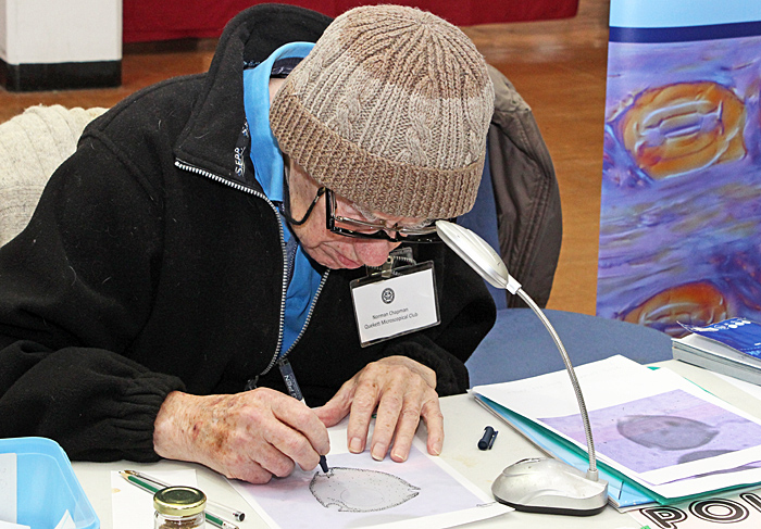 Norman Chapman drawing a pollen grain on tracing paper over a printed photomicrograph
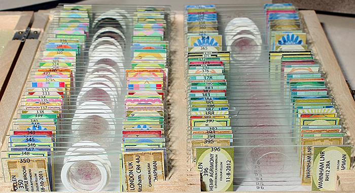 Norman Chapman’s pollen slides
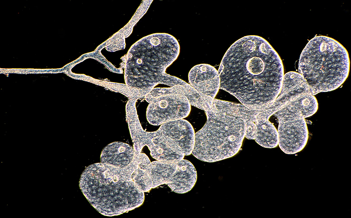
The beauty of pollen grains – a photographic approach
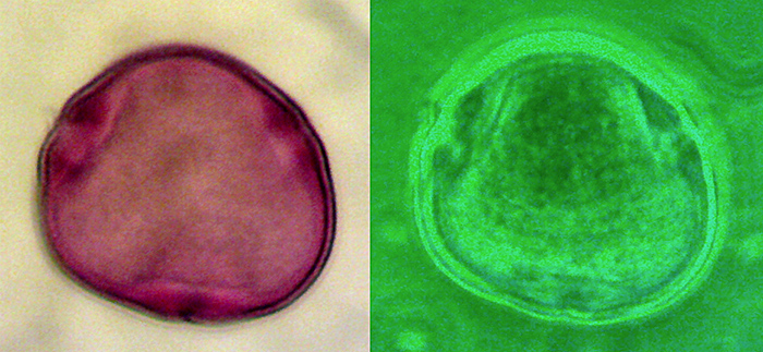
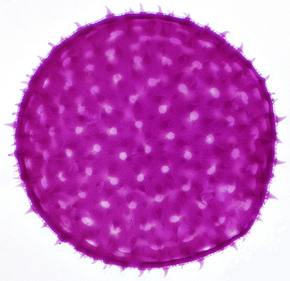 Stained single grain of pollen of mallow (Malva sp.) from a commercial slide by NBS, diameter 150 µm, stained with fuchsin
Olympus SPlan 40× objective, NFK 2.5× photo eyepiece, stack of 12 images in Zerene Stacker
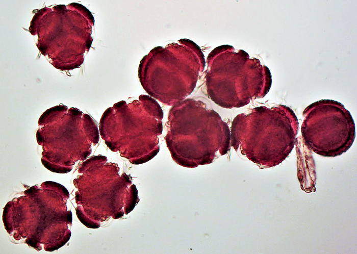
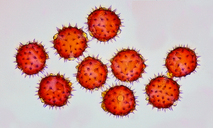 Sunflower pollen: Temporary mount in Calberla’s Solution (DI water, IPA, glycerine, Basic Fuchsin). Leitz SM-Lux, 10× objective, brightfield. 5MP Chinese microscope camera. Stack of 15 images post processed as a focus merge in Affinity Photo. [slide and photo by Gordon Brown]
Using different types of illumination can also reveal details in pollen grains
 Lime pollen grain using brightfield (left) and phase contrast illumination [by John Rhodes]
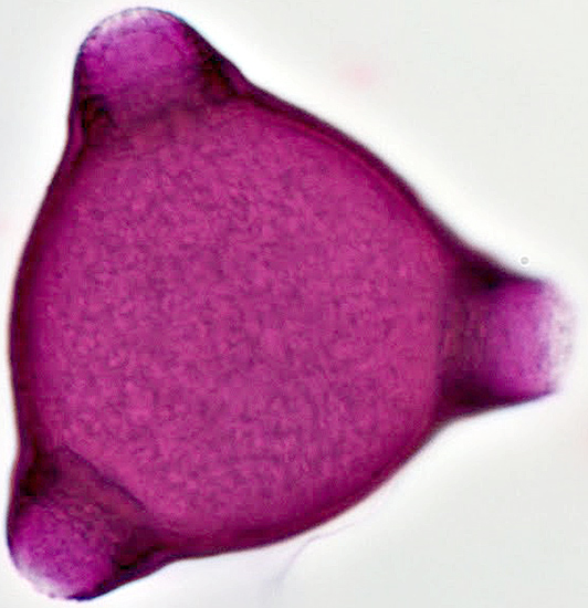 Single pollen grain of rosebay willowherb [slide by David Galliford]
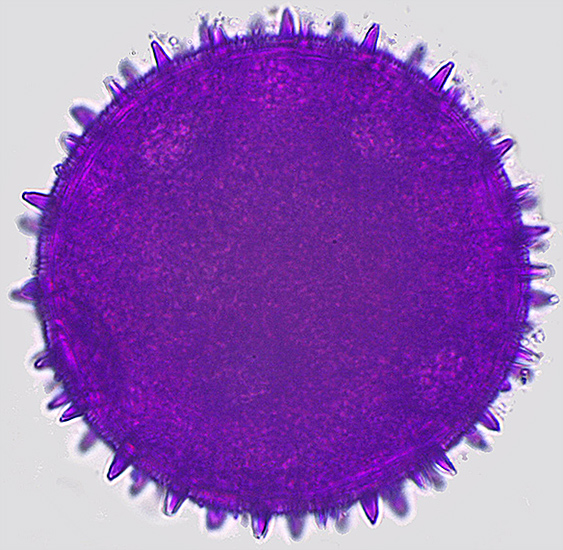
 Pollen of Passiflora [slide by John Blakesley]
 Single pollen grain of butternut squash [slide by David Galliford]
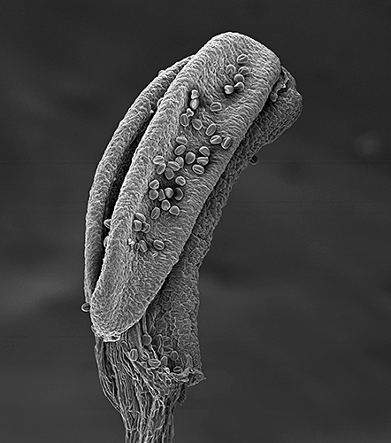 Pollen grains on anther of aconite [SEM by David Spears]
Find the pollen!
Something for the young enthusiast.
Click the arrows to move through the slides. Click the symbol at bottom right for a larger version.
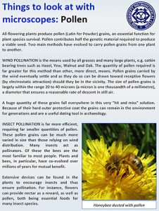 We have an introductory leaflet on using a microscope to observe pollen. This small handout is one of a series aimed at families where parents / grandparents want to encourage children to examine the ‘microworld’.
- Things to look at with microscopes: pollen (PDF: This leaflet is intended to be printed double-sided and then folded to produce a leaflet, and so the page order does not look right when you view it on a computer screen.)
Honeybees
Stereomicroscopes are easy to use and very useful for looking at specimens the size of honeybees, but making a stereo image that can be viewed on a computer screen is not so easy.
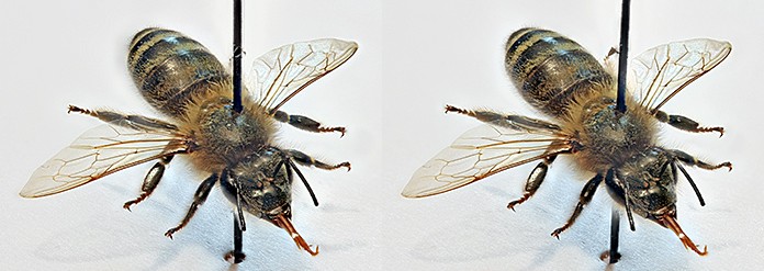
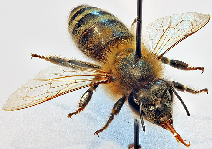 Pinned specimen of a honeybee (Apis mellifera)
Canon EOS 40D with 60 mm EF-S macro lens at f/4.5, stack of 25 images in Zerene Stacker, focus steps using EOS Utility
Some of the image stacking programs that are used to produce macro photos and photomicrographs with good depth of field can also be used to generate images that can be combined to generate stereo images.
 Rocking animation of a honeybee (Apis mellifera), images generated by Zerene Stacker and combined into an animated GIF in Photoshop Elements
Alan Cooper of the Stereoscopic Society recommends StereoPhoto Maker for producing stereoscopic images, so Alan Wood exported 2 images from Zerene Stacker as a stereo pair and then used StereoPhoto Maker to produce images for free viewing and for red/cyan 3-D anaglyph glasses.
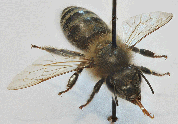
 Stereoscopic pair of images of a honeybee for free viewing
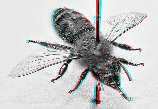 Stereoscopic image of a honeybee for viewing with red/cyan anaglyph glasses
Parasites, pathogens and pests
Honeybees have all sorts of natural enemies, and while light microscopes cannot be used to identify viruses they can be used to observe and identify many other pests, parasites and pathogens.
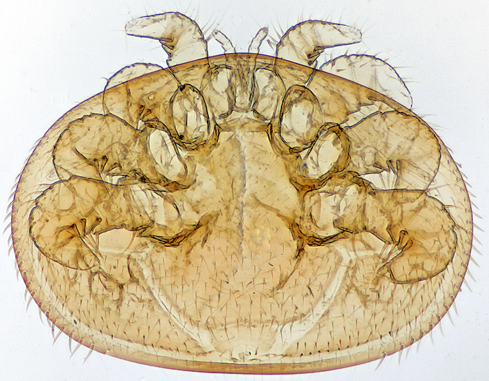 Cleared female Varroa destructor, mite is 1.65 mm wide, slide by L & P Bircham
Olympus SPlan 4× objective, NFK 3.3× photo eyepiece, stack of 22 images in Zerene Stacker
 Adult of Braula coeca (a wingless fly), slide by Peter Sunderland
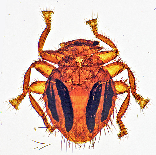
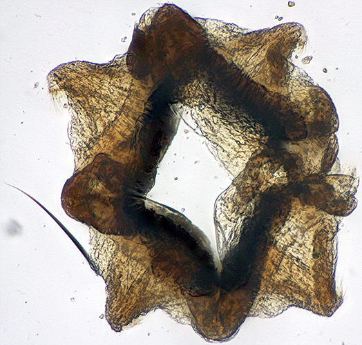
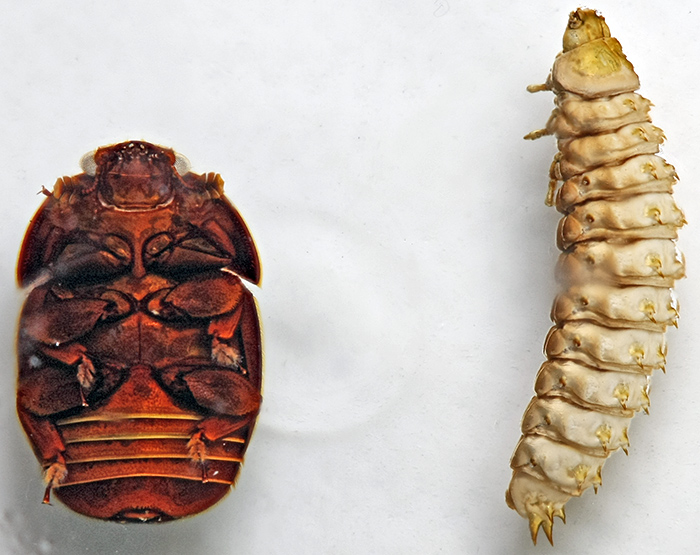 Underside of adult small hive beetle (Aethina tumida) (left) and larva (right); the adult is 5 mm long
Canon EOS 40D with 60 mm EF-S macro lens at f/4.5, stack of 7 images in Zerene Stacker
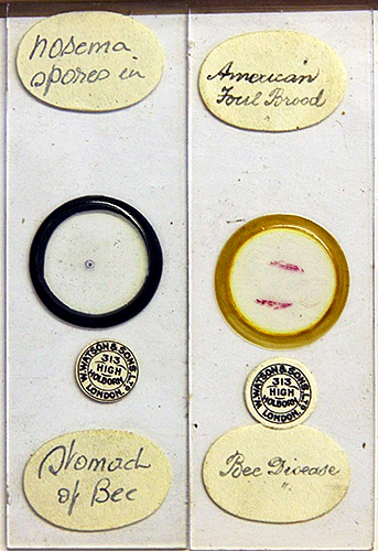 Antique Watson slides of Nosema and American foul brood
Something for the history buffs:
- The Laboratory Diagnosis of Honey-Bee Diseases, by H. A. Dade (PDF)
Anatomy
Even more innovative photography.
 Corbicula (or ‘pollen basket’ and ‘pollen press’) on the hind leg of a honeybee, from a slide made by Dennis Fullwood at Flatford Mill Field Centre in February 2016. Leitz PL ×6 objective and a ×2.5 relay lens, stack of 10 images, photo by Ray Sloss.
 Mouthparts of Apis mellifera, taken with Olympus TG-4 camera on microscope setting, slide and image by John Rhodes
 Head gland of honeybee, dark-ground illumination, 6.3× objective [slide by Brian Norman]
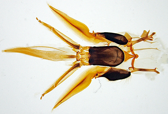
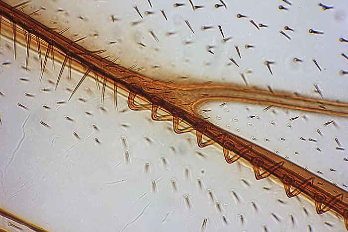 Fore and hind wings of honeybee, 16× objective [slide by John Rhodes]
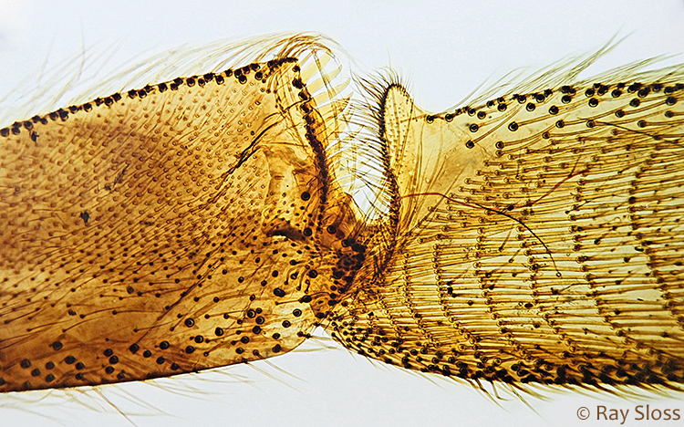
 Proventriculus of honeybee [slide by John Rhodes]
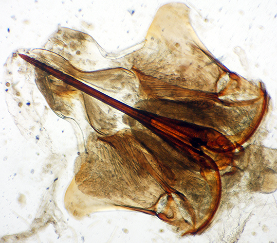 Sting of honeybee, 2.5× objective [slide by John Rhodes]
Something for the history buffs:
- The Malpighian Tubules of the Honey-Bee, by H. A. Dade (PDF)
Explore our website and social media
See how to develop your microscopy and photomicrography talents.
Check our Facebook group to see our members’ current work.
Contact our membership secretary for more information.