

Images from the website
Members of the Quekett have kindly allowed us to use some of their photographs on the Club’s website. The design of the site does not allow us to acknowledge the photos in situ, so they are collected here with details of the photographer, subject and equipment.
Copyright of these photographs is owned by the named photographers.
Gyrinus swimming leg
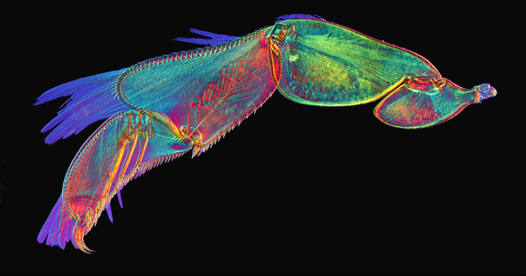
Photograph Copyright © Dr David Linstead. Swimming leg of whirligig beetle (Gyrinus sp.) by polarised light.
Doto sp.
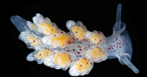
Photograph copyright © James Robson. Nudibranch sea slug (Doto sp.) by dark-ground illumination.
- See more photographs from the 2013 Marine Microscopy Weekend (Quekett members only)
Diaphorodoris luteocincta
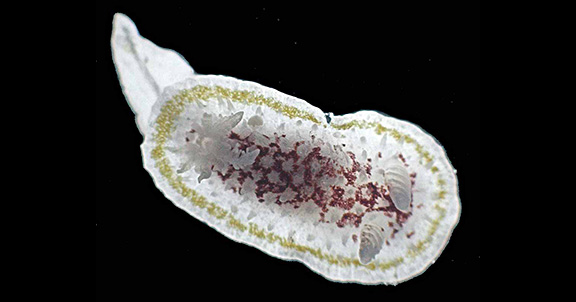
Photograph Copyright © Terry Hope. Nudibranch sea slug (Diaphorodoris luteocincta (M. Sars)) by dark-ground illumination.
- See more photographs from the 2011 Marine Microscopy Weekend (Quekett members only)
Potentilla leaf
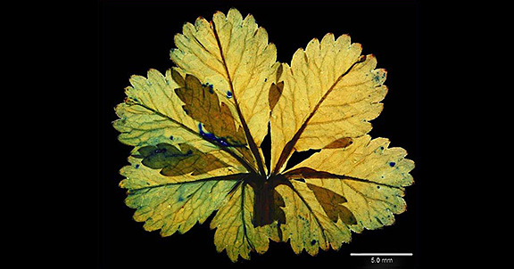
Photograph Copyright © Brian Singleton. Leaf of cinquefoil (Potentilla reptans L.) under dark-ground illumination, Nikon Coolpix CP4500, generic stereomicroscope (maker unknown). Mount by Ernie Ives.
Actinoptychus heliopelta
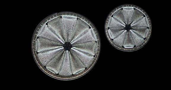
Photograph Copyright © Dr David Linstead. Fossil diatoms (Actinoptychus heliopelta Grunow) by incident light.
Silk moth antenna
Photograph Copyright © Dr David Linstead. Antenna of silk moth, by dark-ground illumination.
Cat tongue
Photograph Copyright © Dr David Linstead. Transverse section of the tongue of a cat, under polarised light.
Culex pipiens larva
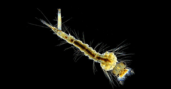
Photograph copyright © Alan Wood. Larva of a mosquito (Culex pipiens L.); slide by T. Gerrard & Co. Olympus Zuiko Auto-Macro 38 mm f/2.8, Olympus Telescopic Auto Tube, Canon EOS 5D Mark II, EOS Utility, dark-ground illumination from an LED ring light, background cleaned up with Photoshop Elements II.
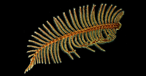
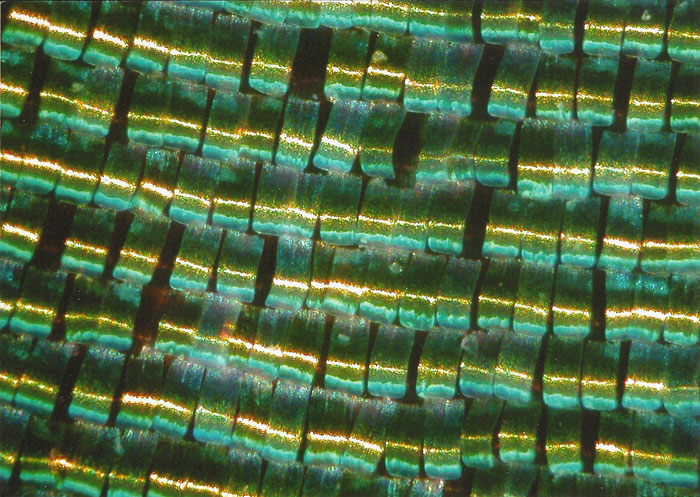
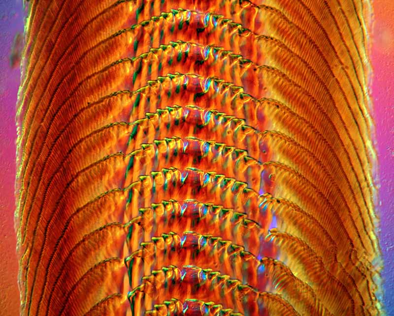
Partridge feather
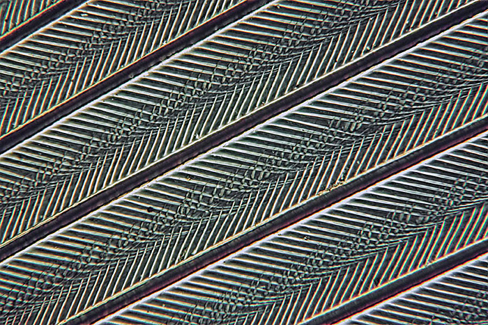
Photograph Copyright © Dr David Linstead. Taken with a 6.3× objective and polarised light. Stack of 20 images.
- Read David’s article on Enhancing depth of field by combining image stacks
Wing scales of Chrysiridia ripheus
Photograph Copyright © Professor Maurice Moss. Iridescent wing scales of a moth, Chrysiridia ripheus (Drury). Nikon Coolpix 4500 camera on a Watson System 70 microscope with ×10 objective. Illuminated with a very oblique light source, set up to light the scales in the direction that they lie along the wing.
- Read Maurice’s article on Scales of Chrysiridia ripheus
Vulture louse
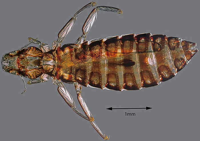
Photograph Copyright © Dr David Linstead. A large louse (~ 4 mm) from a Bengal Vulture; slide made by Charles Collins Junior, who sold slides under the name “Micro-naturalist”. Canon EOS 40D camera on a Nikon Diaphot inverted microscope with LWD 0.55 Phase/DIC condenser. Nikon ×10 Plan DIC objective. A total of 1529 images in 48 stacks. Differential interference contrast (DIC).
- Read David’s articles on Stacking and Stitching
Coryne eximia
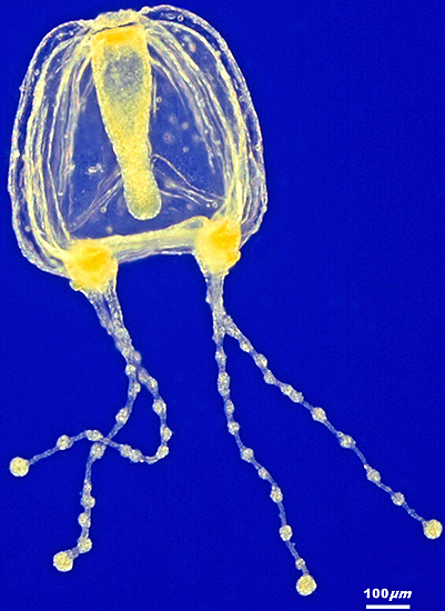
Photograph Copyright © Carel Sartory. A fluid mount of a newly-liberated medusa of Coryne eximia Allman. Stack of 22 images. Rheinberg illumination using a filter with a blue central patch and a yellow outer ring.
- Read Carel’s article on Making Rheinberg illuminations discs
Polyester mesh
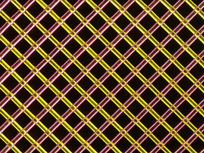
Photograph Copyright © Carel Sartory. Colourless polyester mesh under Rheinberg illumination using a yellow/red quadrant filter with a black central stop.
- Read Carel’s article on Making Rheinberg illumination discs
Diatom Crown
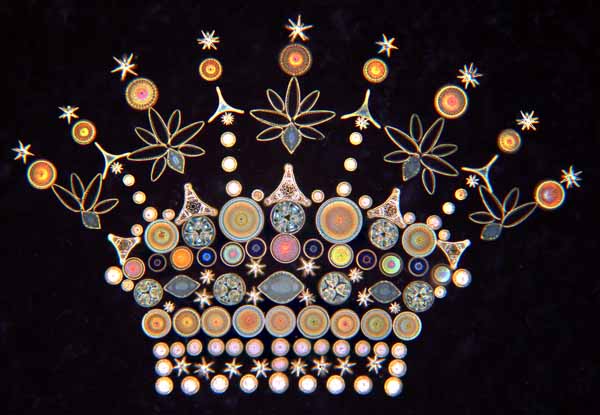
Photograph Copyright © Brian Davidson. 160 Diatoms and sponge spicules arranged as a crown by William Gatrell. The centric forms give a striking diffraction effect under dark-ground illumination.
- Read Brian’s notes on this slide (Quekett members only)
Palate of Trochus cinereus
Photograph Copyright © Brian Davidson. Palate of a marine snail, Trochus cinereus, from a slide made by T. T. Hennah in 1864. Olympus BH-2 microscope with a 16mm objective, using differential interference contrast (DIC).
- Read Brian’s notes on this slide (Quekett members only)
Daphnia pulex
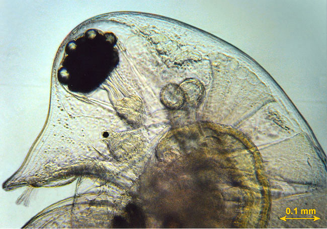
Photograph Copyright © Graham Matthews. Live wet mount of the water-flea Daphnia pulex (Linnaeus).
Insect in Drosera
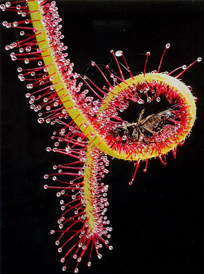
Photograph Copyright © Duncan Edmonds. An insect trapped by the insectivorous sundew plant Drosera.
Eggs of cabbage white butterfly
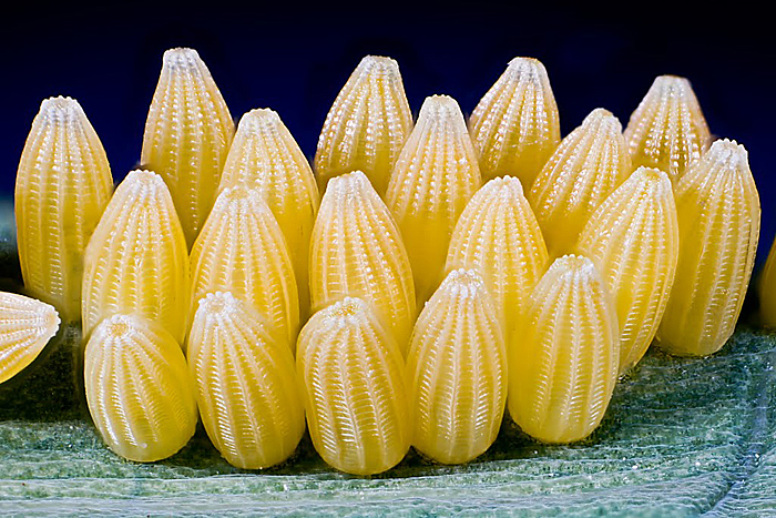
Photograph Copyright © Alan Casperd. Eggs of Pieris brassicae (Linnaeus).
Christmas tree arrangement of diatoms
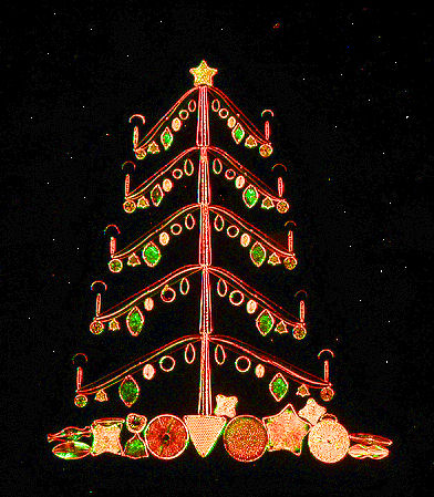
Photograph Copyright © Tony Saunders-Davies. Slide of arranged diatoms by Klaus Kemp. Rheinberg illumination.