

Barnard Awards 2023 (Artistic)
The judge, Alan Edwards ARPS, considered the photographs before they were shown at the Annual Exhibition of Microscopy at Potters Bar on Saturday 14th October 2023, and had the difficult job of deciding which of the ones from members of the Quekett, the Iceni Microscopy Study Group and the Postal Microscopical Society were of a sufficiently high standard to deserve a certificate.
He awarded a certificate to David Furness (Housefly recoloured), Mike Gibson (Looking at you), David Linstead (Section of Pinna shell), Sandie Pearce (Kojic Butterfly) and James Rider (Salicine crystal slide).
Quekett members can see larger images and the judge’s comments in the Photography showcase.
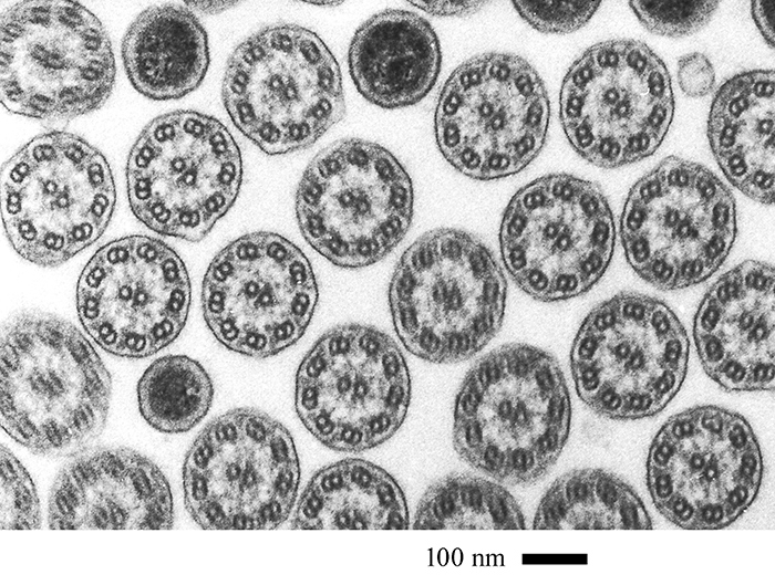 David Furness: Field of cilia
Field of cilia in transverse section obtained using my JEOL S100 transmission electron microscope. The project is about the structure and organisation of ciliates and is ongoing. The sample is a resin block made in 1981 containing rumen ciliates. These were sectioned at 90 nm thickness, stained with uranyl acetate and lead citrate, and examined in the TEM. Images were recorded onto Acros Neopan 100 35 mm film, developed and digitally scanned using a Canon flatbed scanner. The highly symmetrical cilia can be seen with their 9+2 arrangement of microtubules and associated proteins. In between are cylindrical bacteria (the dark profiles) also in transverse section that live between the animal’s cilia.
Processing the block took 3 days, cutting sections a week and staining 30 minutes. Observation time was 2–3 h.
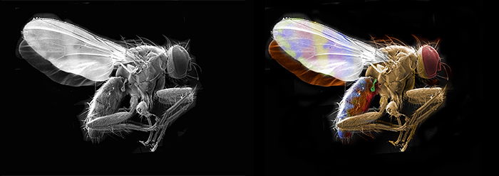 David Furness: Housefly recoloured (Barnard Award)
Dead, air-dried housefly collected from window ledge in my garage. Using a sticky carbon pad, this was stuck to an aluminium stub suitable for insertion into the microscope. Sputter-coated with a thin layer of gold and placed for imaging. Five images of the fly were collected at an observing magnification of 50× and montaged together using Adobe Photoshop 7 software. A copy of the grey-scale image was false-coloured using approximate colourings for a live fly.
JEOL T300 scanning electron microscope.
 David Linstead: Section of Pinna shell (Barnard Award)
Section of the shell of Pinna nobilis, whose common name is the noble pen shell or fan mussel, a large species of Mediterranean clam, a marine bivalve mollusc.
Mounted slide from the Victorian/Edwardian period, sold by Watson.
Nikon Diaphot inverted microscope, Zeiss Jena ×10 plan apo objective, oblique polarised light with Cellophane retarder. Canon EOS 5D Mark II camera directly mounted to camera port. Image not stacked or stitched.
 David Linstead: Section of fossilised shark tooth
Thin transverse section of a fossil shark tooth. Slide mounted specimen from the Victorian/Edwardian era. Mounted and sold by C. M. Topping.
Imaged with a Nikon Diaphot inverted microscope. DIC illumination with a Nikon ×10 DIC objective and a Nikon variable retarder. Image not stacked or stitched.
 James Rider: Salicine crystal slide (Barnard Award)
Slide by J. G. Bradbury, paper-covered and appears to be vintage.
Olympus BHS with 4× SPlanApo objective, Achromat Aplanat condenser and crossed polarizers.
Full-frame Canon EOS 6D Mark II camera, ISO 100.
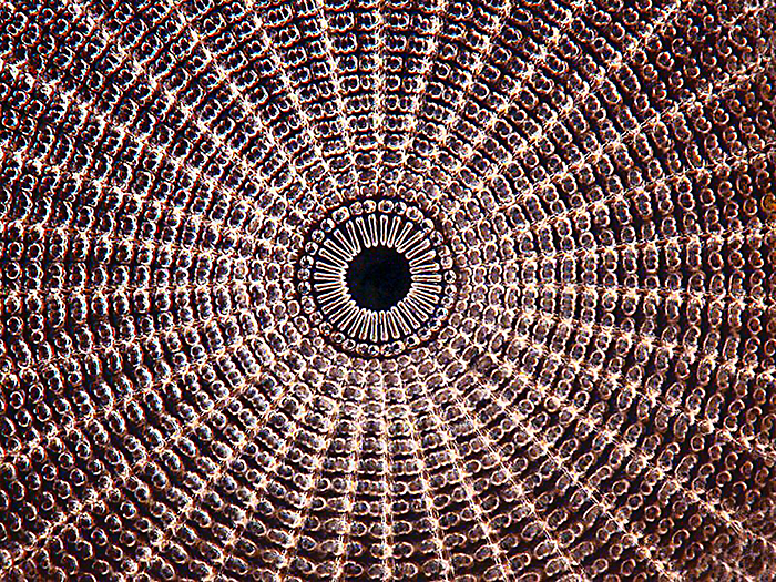 Les Franchi: Diatom view
Diatom – Arachnoidiscus ehrenbergii from an arranged group by Herbert Potter.
Dark-field view, ×40 objective ×10 ocular.
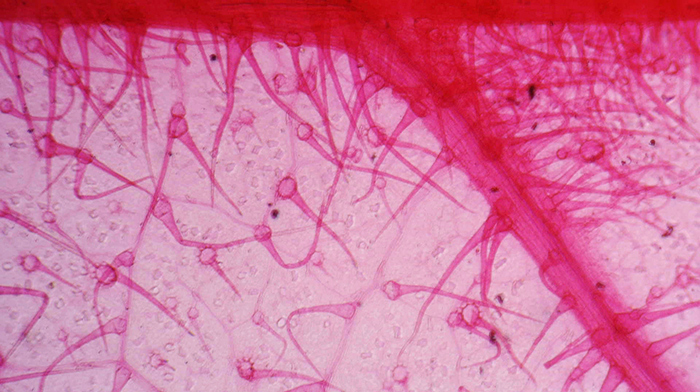 Lisa Ashby: Elm leaf
Elm leaf stained with safranin and mounted by Ernie Ives.
Watson Bactil 60 trinocular, ×10 Para objective, transmitted light.
Chinese USB camera, capture through Micam software.
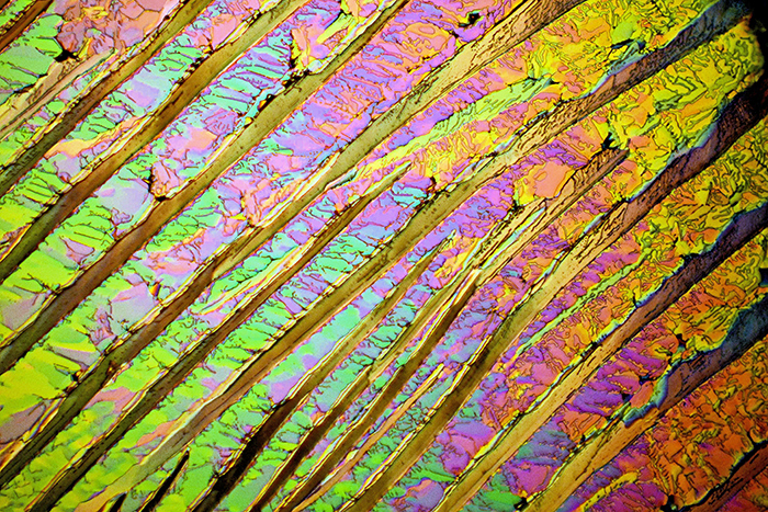
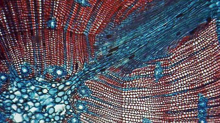 Lisa Ashby: Pinus sylvestris
Transverse section of Pinus sylvestris mounted in 1935 by F. Weait. Mounted in xylol, stained with safranin and clove oil.
Watson Bactil 60 trinocular, ×10 Para objective, transmitted light.
Chinese USB camera, capture through Micam software.
 Mike Gibson: Ascorbic acid crystals
Crystals viewed with polarized light on a Wild M20 microscope with ×10 objective.
Camera: Sony Cyber-shot DSC-W35, 0.2 sec exposure, f/4, ISO 100.
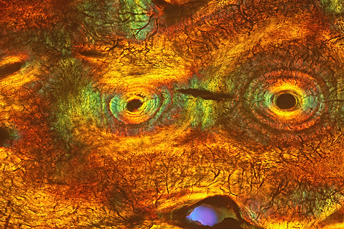
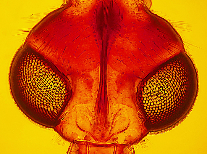 Mike Gibson: “Looking at You” through compound eyes of a fly (Barnard Award)
Photograph from a prepared Biosil slide by the late John Wells.
Taken with a Sony compact Cyber-shot DSC-W35 camera at a shutter speed of 1/100s, f/11, ISO 100.
Bright-field illumination with addition of Rheinberg filter. Microscope used: Wild M20.
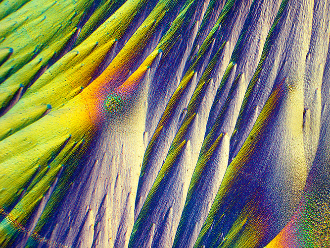
 Sam Medworth: Naphthalene crystals
Cooke, Troughton & Simms petrological microscope, with CTS ×10 objective and unnamed ×10 projection eyepiece.
Nikon D7100 camera with extension tubes to exclude extraneous light, but no camera lens, tethered via USB to a laptop with Nikon Capture NX2. Saturation and contrast adjusted in software.
I melted a piece of mothball on a slide and covered it with a coverslip, then watched it under the microscope while it started to solidify. Under crossed Nicols, the most beautiful colours and patterns appeared.
 Sandie Pearce: Kojic Butterfly (Barnard Award)
Crystals formed from kojic acid, a skin treatment chemical, heated on a slide and imaged with polarised light.
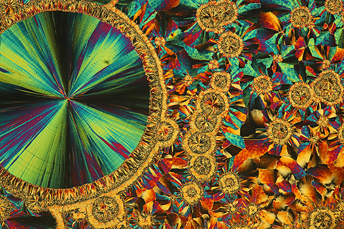
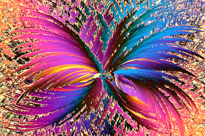
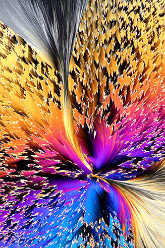 Sandie Pearce: Kojic Whirlwind
Kojic acid, a skin treatment chemical derived from the fermentation process of rice for use in the manufacturing of sake (a Japanese rice wine). Kojic acid is used in cosmetics and in the food industry as a preservative.
I heated the kojic acid to 155°C on a slide until melted, and imaged under polarised light with a full-frame camera mounted on a Nikon E400 microscope.
 Thomas Consi: Termite gut protist – Trichonympha
Image of a living Trichonympha from a termite gut. Wet-mount.
Microscope: Olympus BH-2, probably 10× or 20× objective, differential interference contrast (DIC).
Camera: Canon EOS 80D digital SLR.