

Workshop on epi-illumination
Saturday 1st April 2023
Quekett members can watch a video recording of this workshop.
Epi-illumination involves light reflected from the specimen, and is the normal illumination in metallurgical microscopy and the most common illumination used with stereomicroscopes. Reflections of the light source can be a problem, but can often be reduced using two polarisers or diffused illumination.
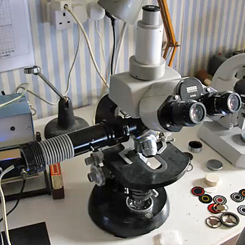
Paul Smith introduced the workshop, describing the principles of epi-illumination and using two small Lomo compound microscopes fitted with epi-illuminators from the Club’s collection of gadgets. They have a male RMS thread at the top and a female RMS thread at the bottom, with coverslips, semi-silvered mirrors or prisms to reflect the light from the lamp down through the objective onto the specimen. The original light sources were not available, so Paul used Ikea Jansjö lamps instead, held in place with sticky tape. He used a variety of specimens, including brown mustard seeds (which have indentations similar to those on golf balls), poppy seeds and coins.
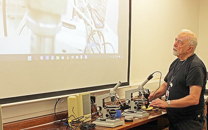 Paul Smith
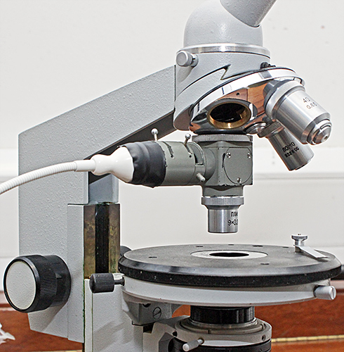 Lomo microscope with Beck epi-illumination adapter
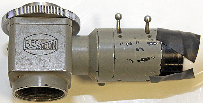 Beck epi-illumination adapter
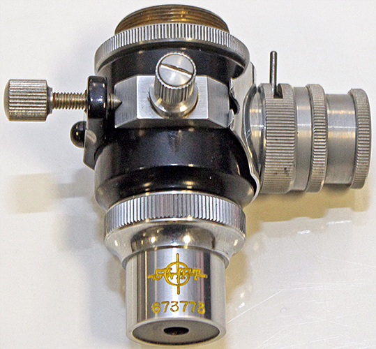 Unbranded epi-illumination adapter
Pam Hamer showed the Cooke Universal Epi-illuminator from a Cooke M35 microscope, which is more elaborate than the Club’s ones and can produce bright-field and dark-field epi-illumination.
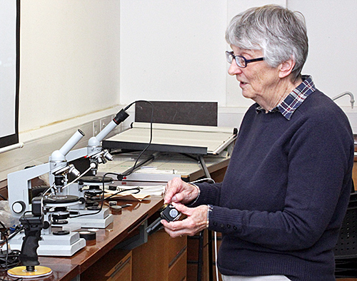 Pam Hamer
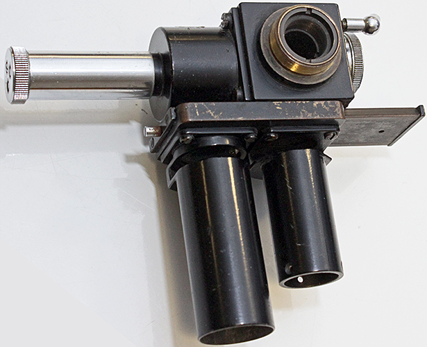 Cooke Universal Epi-illuminator
Cooke Universal Epi-illuminator
(Click the arrow keys to move through the slides, click the bottom right icon for a larger version)
Pam also showed how to use half of a table-tennis ball to produce diffuse epi-illumination with low-power objectives. For illumination,she uses two of the USB-powered Ikea Jansjö lamps that are still available from Ikea.
Table-tennis ball epi-illuminator
(Click the bottom right icon for a larger version)
Graham Matthews used the Club’s Prior trinocular stereomicroscope to show pharmaceutical capsules and tablets that he had cut in half. The simple top light on the microscope worked well with these specimens.
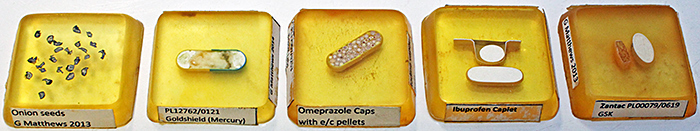 Sectioned pharmaceuticals
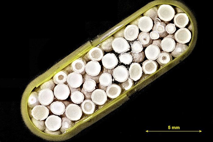 Omeprazole capsule with enteric coated pellets [by Graham Matthews]
Danny Ferri used the stereomicroscope to show us some highly-reflective metal swarf, a thin section of platinum ore and Rizla rolling paper.
Tony Pattinson (via Zoom) showed us a 2764HS EPROM from 1983, looking through its programming or erase window using his Vickers M41 microscope with a reflected-light attachment.
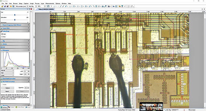 2746HS EPROM
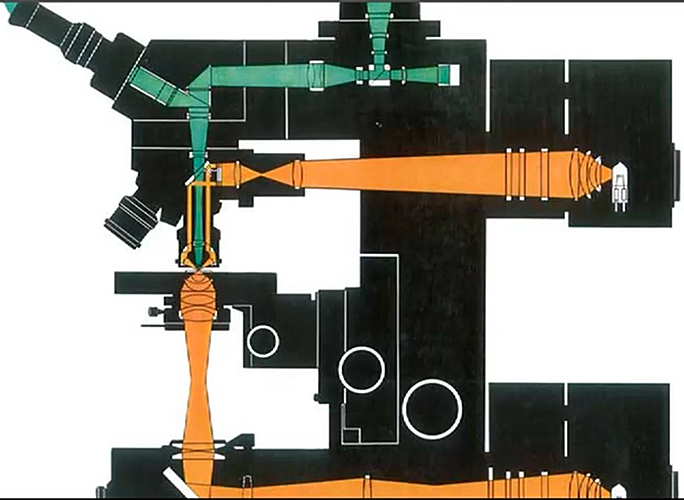 Reflected light path in Vickers M41 microscope
Graham Matthews showed us a photograph of his Zeiss GFL microscope with a reflected-light attachment. The attachment also fits Zeiss Standard microscopes, but the GFL allows taller specimens to be observed.
 Zeiss GFL microscope with reflected-light attachment [by Graham Matthews]
The eyepiece camera had been playing up, giving everything an orange colour, and then failed completely. Alan’s specimens could not be seen by the people participating via Zoom, but he provided some photographs for the report.
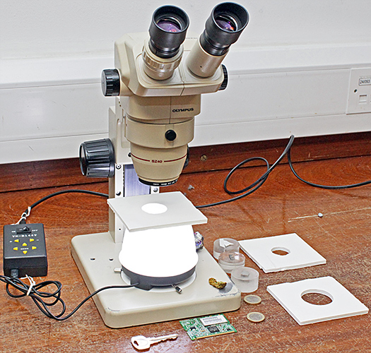
Alan Wood brought his Olympus SZ4045 stereomicroscope with a home-made shadowless, diffused illuminator based on a 144-LED ring-light that would normally be attached to the microscope. He usually uses it with biological specimens, but for the workshop he showed how well it works with metallurgical samples, crystals, coins, a locket and a printed circuit board.
 Alan Wood’s demonstration
The LED ring-light is placed upside down on the stage plate. Inside the ring-light there can be a disk of Plastazote for pinned insects, or a disk of white, black or coloured paper as a background. A roll of white paper keeps direct light from the LEDs away from the objectives. A plastic pudding basin with its base removed sits just outside the ring of LEDs, reflects light onto the roll of paper, and supports a piece of white foam board. The foam board reflects some of the light down into crevices in the specimens. Alan has three foam boards with different sizes of holes for use with different microscope magnifications or with a camera plus macro lens.
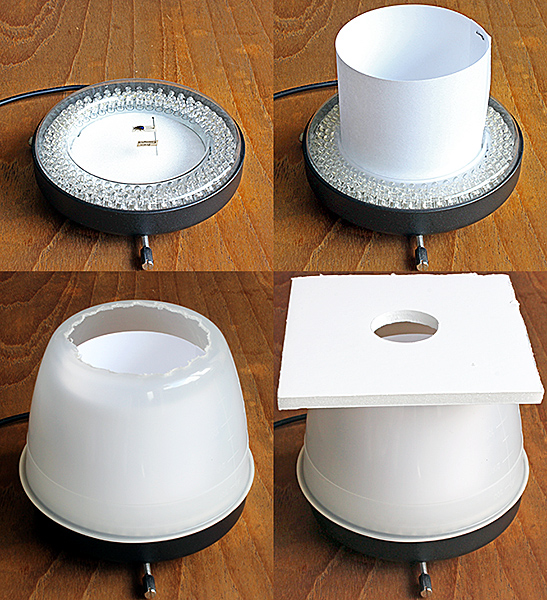 Shadowless illuminator, showing (top left) the inverted ring-light, (top right) ring of white paper, (bottom left) inverted bowl with base removed, and (bottom right) the complete illuminator.
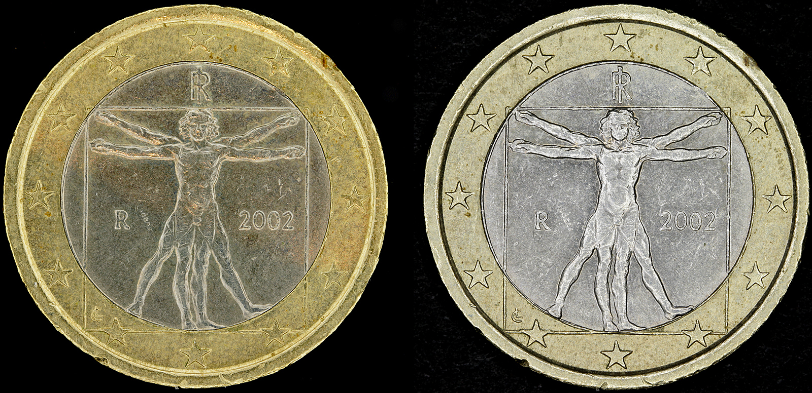 1 euro coin (left: shadowless illuminator, right: LED ring-light)
Click the image to see a larger version
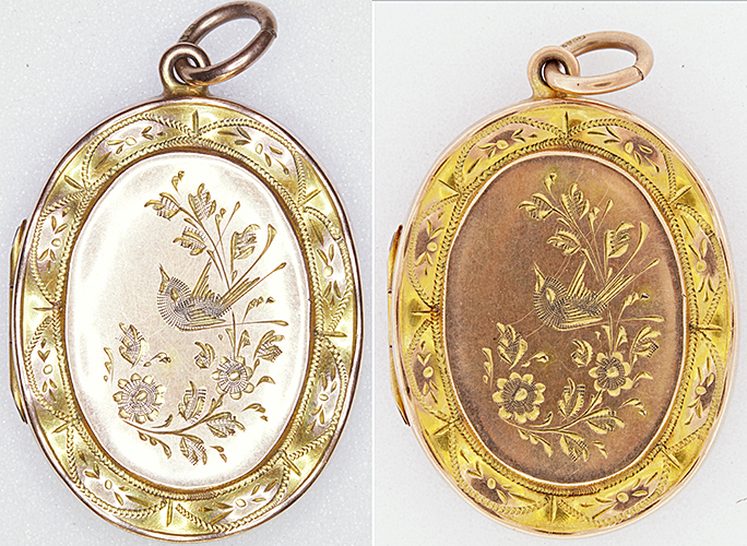 Locket (left: LED ring-light, right: shadowless illuminator)
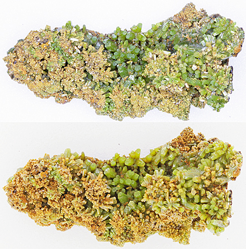 Pyromorphite (top: LED ring-light, bottom:shadowless illuminator)
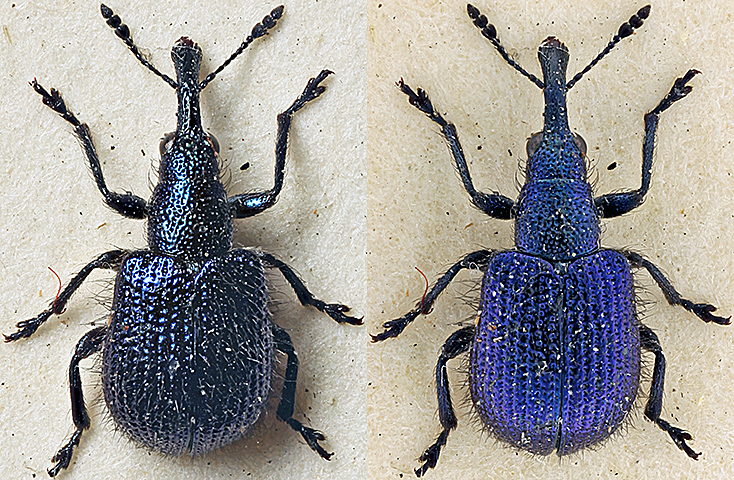 Small weevil (left: desk lamp, right: shadowless illuminator)
Alan had intended to bring his Olympus BHMJ-N metallurgical microscope with its reflected bright-field and dark-field illumination and matching objectives, but it was too heavy so he provided some photographs for the report instead.
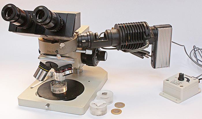 Olympus BHMJ-N metallurgical microscope
The illuminator has two settings, one for bright-field to send light to the specimen through the lenses in the objective, the other for dark-field to send light to the specimen through a passage around the lenses in the objective. The special nosepiece has a central channel to send light and images through the lenses, and a second outer channel to send light around the lenses. The objectives have a 25 mm thread (wider than RMS) to provide room for the outer channel, and are corrected for a 210 mm tube length (instead of the normal 160 mm) to allow for the height of the illuminator.
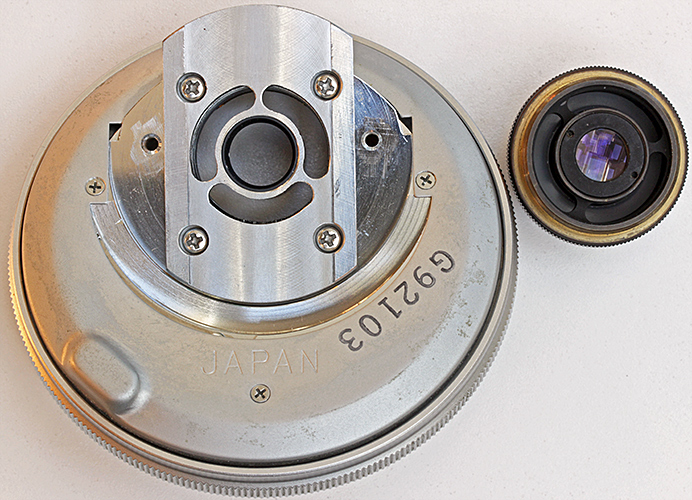 Olympus BH-NRE nosepiece and Neo 5× objective
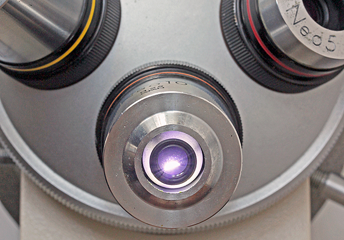 Olympus Neo 10× objective showing dark-field ring of light
Reflections and flare can be problems, so this type of microscope is usually supplied with two polarisers. Through the microscope, the depth of field is extremely shallow (around 0.05 mm for the 5× objective, even less for higher powers), but image stacking allows photographs with reasonable depth of field to be produced.
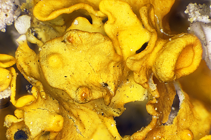 Stacked image of yellow lichen (Olympus Neo 5× objective, FK 2.5× photo eyepiece, stack of 44 images, 1.75 mm depth of field)
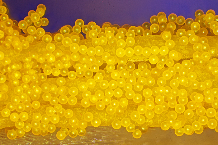 Stacked image of crocus pollen on stamen (Olympus Neo 5× objective, FK 2.5× photo eyepiece, stack of 42 images, 1.7 mm depth of field)
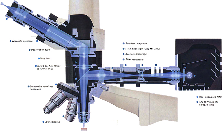 Light paths in Olympus BH-2 metallurgical microscopes
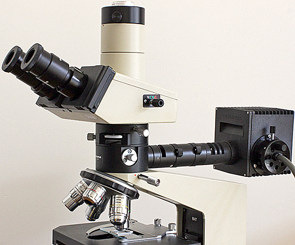 Olympus BH-2 metallurgical microscope
Report and most photographs by Alan Wood