

Warnham Local Nature Reserve excursion
Saturday 5th August 2023
This was the twelfth Quekett excursion to Warnham Local Nature Reserve on the outskirts of Horsham, West Sussex, hosted by the Friends of Warnham Local Nature Reserve, and organised by Graham Matthews. Since our last visit in 2019, a new pond and a new reed bed have been created near the entrance, and a Discovery Hub has been built on the edge of the millpond.
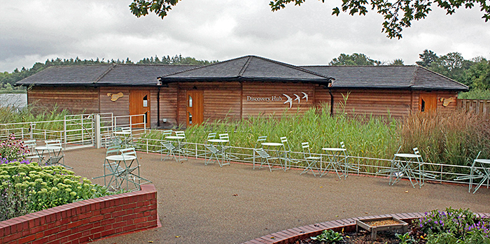 Discovery Hub
There are no microscopes in the Hub, but it does have a magnifier on a flexible arm showing images on a LED screen:
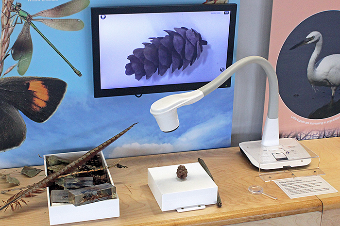 Magnifier in the Hub
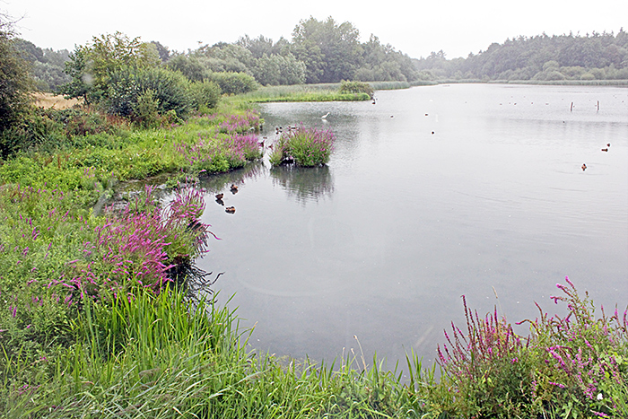 Millpond (viewed from the Discovery Hub)
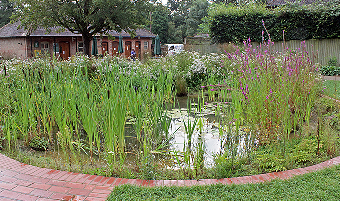 New pond
In addition to the new pond and reed bed, the Reserve includes a large millpond, 3 dipping ponds and an extensive board walk that provides access to a variety of streams, ditches and marshy areas.
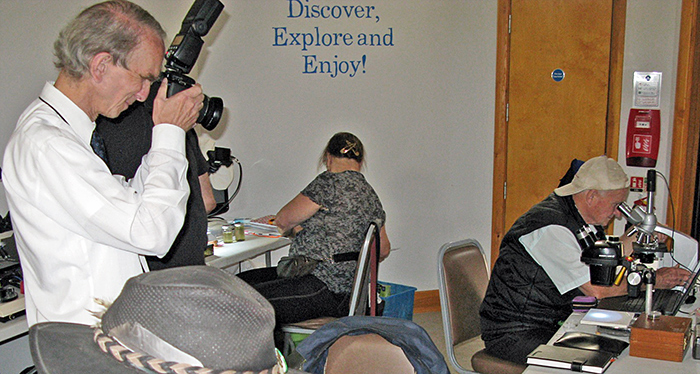
We set up our microscopes, cameras and computers in the Discovery Hub, and we had permission to use our nets and bottles to collect specimens to examine under our microscopes.
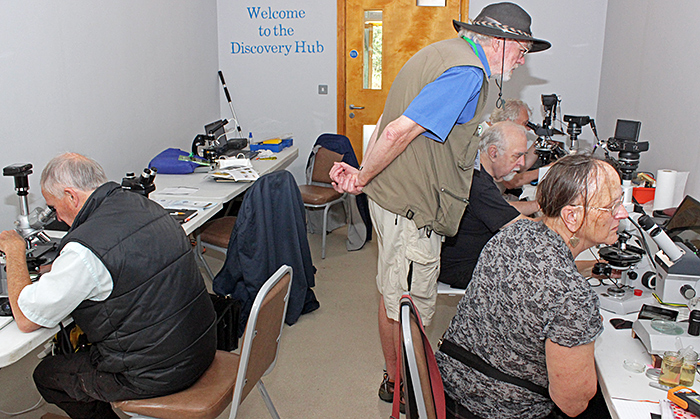 Quekett members in the Discovery Hub
Storm Antoni made it difficult to go out collecting, but we did manage to get some samples from the new pond and from the dipping ponds.
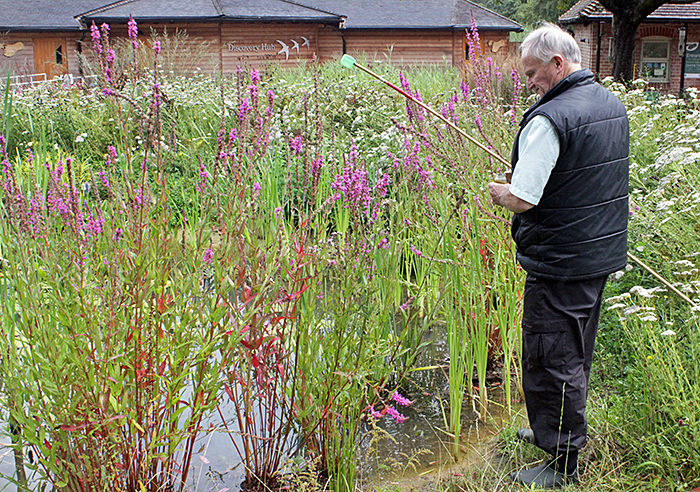 Mark Berry collecting from the new pond
There had been lots of rain recently, so there was plenty of water in the dipping ponds this year.
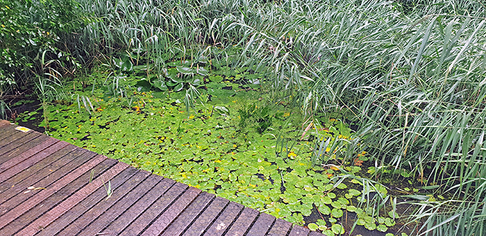 Dipping pond [by Graham Matthews]
Jacky McPherson used a small plastic bottle attached with gaffer tape to a telescopic golf ball retriever.
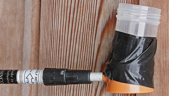 Plastic bottle on golf ball retriever
Mark Berry’s equipment included a miniature plankton net on a fishing rod.
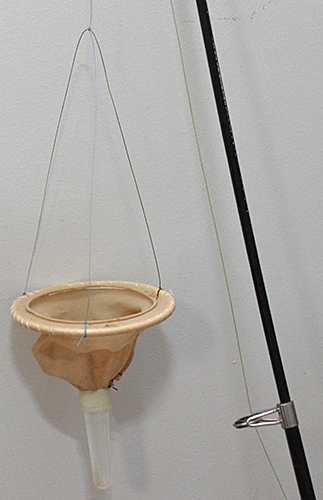 Miniature plankton net
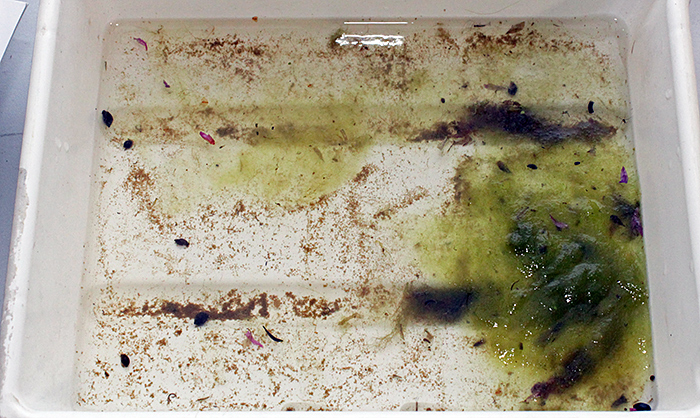 Sample from the new pond
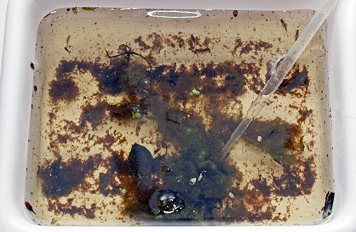 Sample from the dipping ponds
We could immediately see water-fleas and mayfly nymphs, but we needed our microscopes to observe most of the specimens.
Graham Matthews brought his trinocular Leitz Dialux compound microscope with a rotating stage and DIC. Illumination was via a randomised Y-shaped fibre-optic cable with electronic flash on one arm and continuous light on the other. For taking photographs, he used an afocal arrangement with a Canon EOS 500D digital SLR sending images via USB to DSLR Remote Pro on his laptop computer. He used Helicon Focus for stacking. Graham has provided several photomicrographs of his specimens.
 Graham Matthews
Graham has used some of his photomicrographs and macro photographs to produce a booklet on the freshwater life in the Reserve:
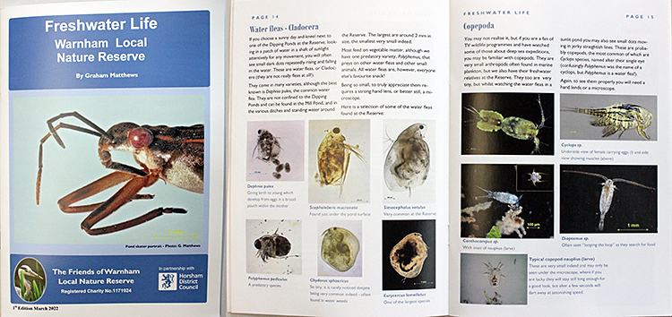 Freshwater Life: Warnham Local Nature Reserve
Jacky McPherson brought her Zeiss Stemi stereomicroscope that she has fitted with LED incident and transmitted light. She also borrowed Graham’s trinocular Zeiss Standard compound microscope. For taking photographs, she used an afocal arrangement with a Canon EOS M3 mirrorless camera fitted with a Minolta Rokkor 45 mm f/2 lens, viewing the images via HDMI on an old flat-screen television. Jacky has provided a video of some of her specimens.
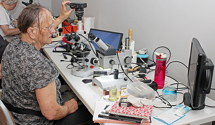 Jacky McPherson
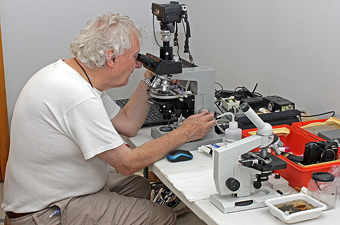
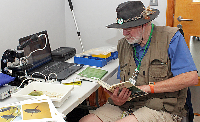
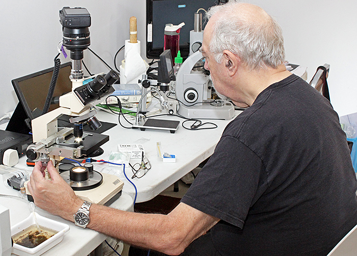
Mark Berry brought his trinocular Reichert microscope. He bought the bare stand at Microscopium in 2022 and has since added a Reichert trinocular head, B&L eyepieces, G&S objectives, a Vickers substage and LED lighting. He also brought a Bausch & Lomb StereoZoom microscope on a makeshift stand with an LED torch for illumination. The specimens that Mark found included Metopus (2 or 3 species), gastrotrichs (2 species), amoebae, testate amoebae (3 species), Arcella, Actinophrys, Difflugia, Daphnia, several species of euglenoid (including Phacus), desmids (several species including Cosmarium), diatoms (several species including large motile form nearly 0.3 mm), rotifers (several species including bdelloid forms), Pediastrum, Merismopedia and Vorticella. To take photographs, he used an eyepiece camera sending images via USB to his laptop computer, and a small Canon point-and-shoot camera. Mark has provided several photomicrographs of his specimens.
 Mark Berry
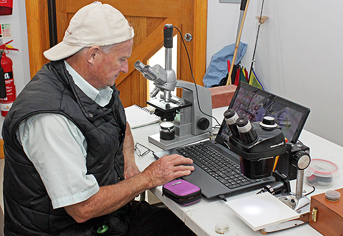
Neil Henry brought his monocular PZO compound microscope fitted with a YW5.0M eyepiece camera, sending images via USB to ToupView software on his laptop computer.
 Neil Henry
Paul Smith brought his trinocular Swift M1000-D compound microscope. For taking photographs, he used an afocal arrangement with a Canon EOS M3 and a Canon EF 40 mm lens, viewing the images via HDMI on a portable monitor. Paul has provided some photographs of his specimens.
 Paul Smith
Alan Wood saw that the weather forecast was for heavy rain all day, so he decided not to carry a microscope as well as a camera for the 25-minute walk from Horsham Bus Station
 Alan Wood [by Graham Matthews]
Our location in the Discovery Hub was not obvious to visitors to the Reserve, so our only visitor was the Countryside Warden, Jake Everitt. Graham has been helping Jake acquire the components for a good microscope, and he now has a trinocular Leitz Dialux, ready to attach a camera.
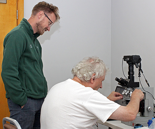 Jake Everitt and Graham Matthews
We used a variety of books and keys to identify specimens, including:
- Collins Field Guide to Freshwater Life by R. Fitter & R. Manuel
- Ponds and small lakes: Microorganisms and freshwater ecology, by Brian Moss
- Small Freshwater Creatures, by Lars-Henrik Olsen, Jakob Sunesen & Bente Vita Pedersen
Specimens
Graham Matthews’ specimens
 Gastrotrich: Neogossea sp.?, 25× objective
 Gastrotrich: Neogossea sp.?, 25× objective
 Gastrotrich: Chaetonotida, Chaetonotus sp. maybe?, 25× objective
 Rotifer: Anuraeopsis fissa? 25× objective
 Rotifer: Anuraeopsis fissa? 25× objective
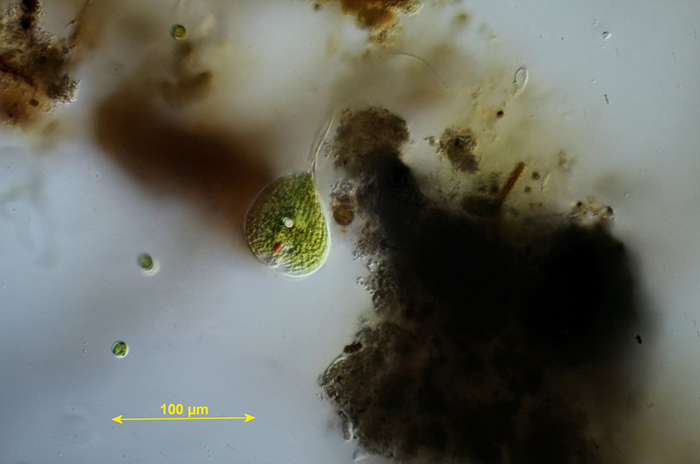
 Euglenoid: Trachelomonas sp. ? 25× objective
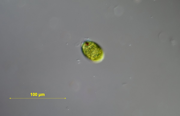
 Euglenoid: Phacus sp., 25× objective
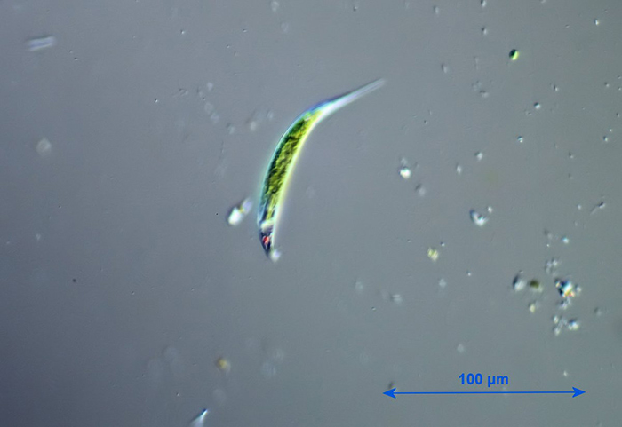
 Euglenoid: Euglena sp., 25× objective
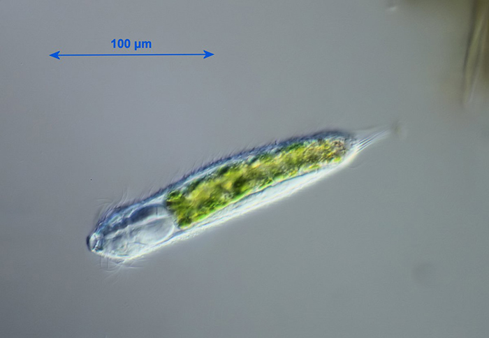
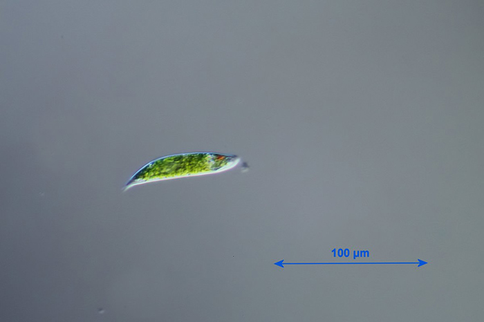
 Euglenoid: Lepocinclis acus, 25× objective
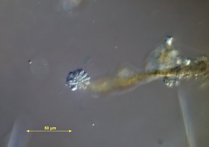 Chromulinaceae: Anthophysa sp., 40× objective
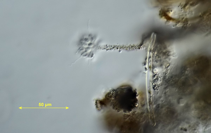 Chromulinaceae: Anthophysa sp., 40× objective
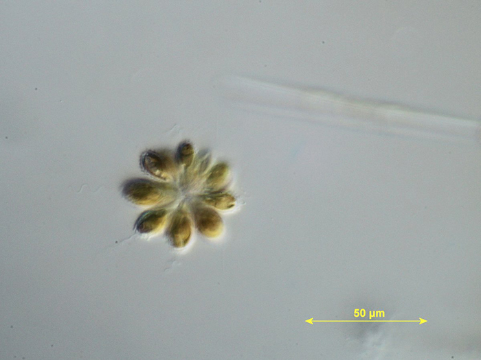 Chrysophyceae: Synura sp., 40× objective
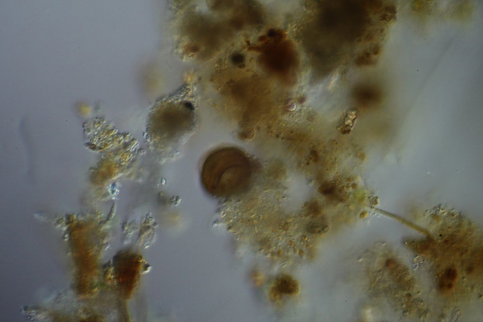 Test of Arcella sp., 25× objective
 Test of Difflugia sp. (?), 25× objective
 Flagellate: Peranema sp., 25× objective
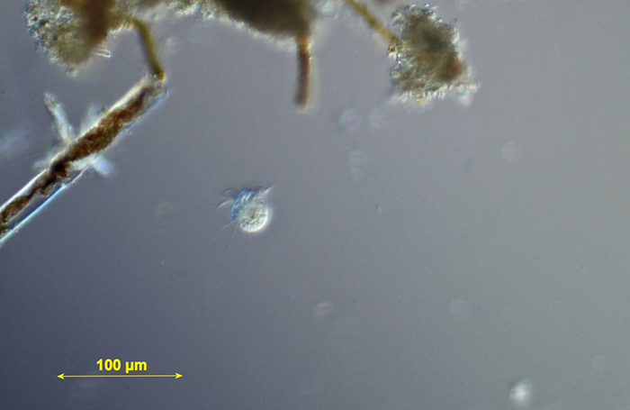
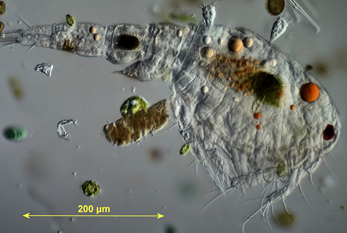 Cyclopian copepod with epizootic peritrichs, 25× objective (including the green epizootic flagellate Colacium sp.)
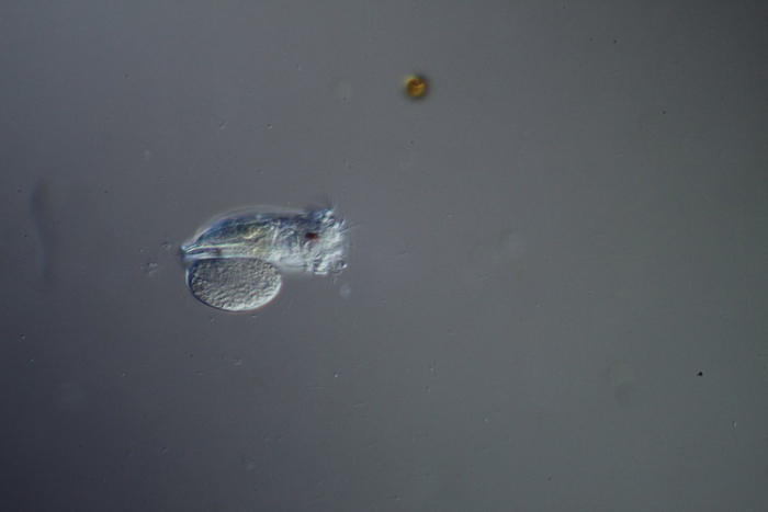
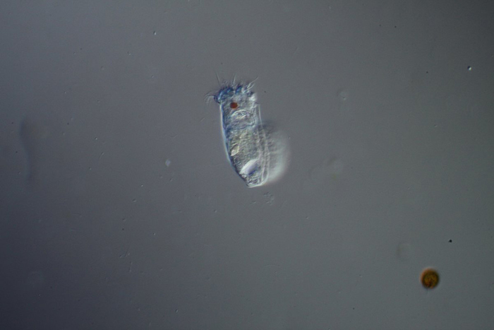
 Cladocera, Chydoridae: Chydorus sphaericus, 25× objective
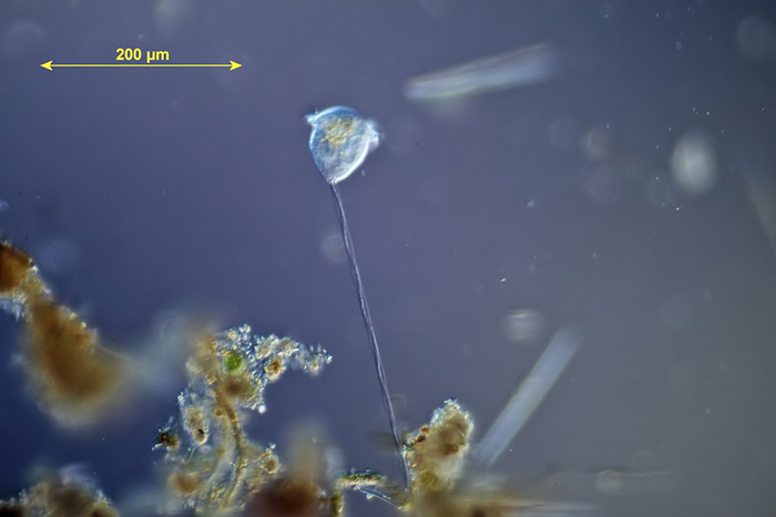 Peritrich ciliate: Vorticella sp., 16× objective
 Ciliate: Askenasia sp. or Halteria sp., 16× objective
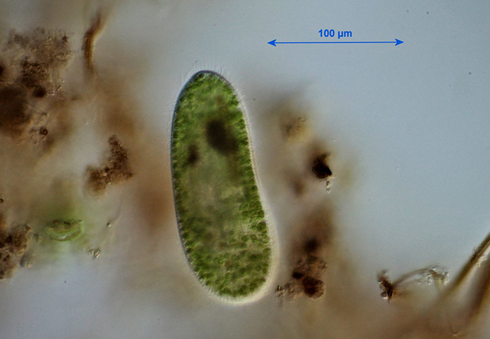 Ciliate: Paramecium bursaria (?), 25× objective
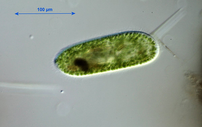 Ciliate: Paramecium bursaria (?), 25× objective
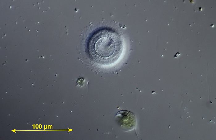 Ciliate: Trichodina sp., 25× objective
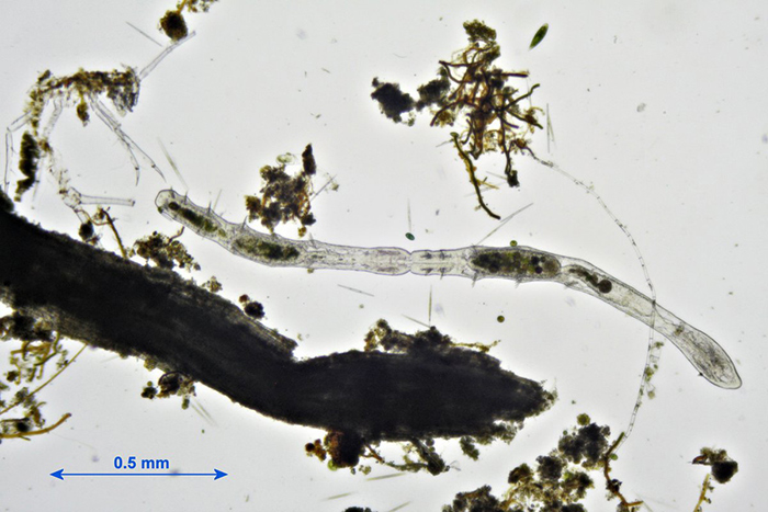 Annelida, Naididae: Chaetogaster sp., 5× objective
Jacky McPherson’s specimens
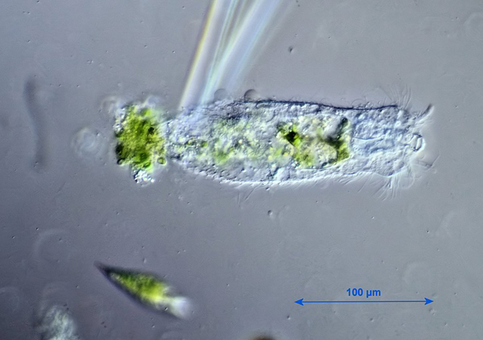
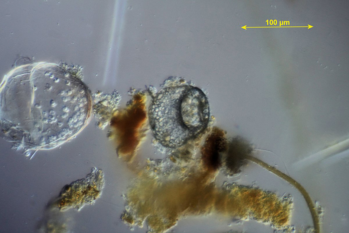
A composite dipping from all three ponds yielded an object of interest which Jacky videoed and has yet to identify. At first it was suggested that the squirming object might be Petalomonas sp., but she could not see any keels and the transformation from a writhing ring (somewhat like an ouroboros) to a long flagellate (flagella visible in life and vaguely shown in the video) did not seem to fit the description. If anyone can clarify what this is, Jacky would love to know. Jacky extracted this image from the video, showing the unidentified flagellate and Synura sp.
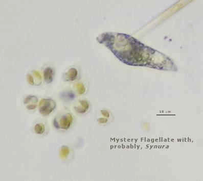 Unidentified flagellate and Synura sp.
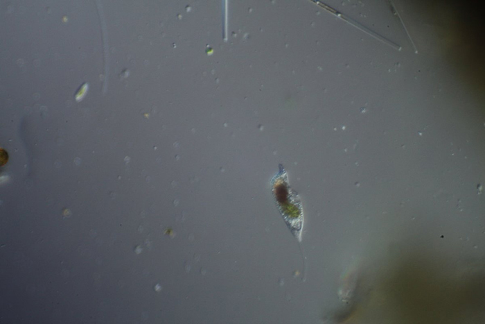
The straight, long, thin entities may well be diatoms, or perhaps Lepocinclis sp., Cyclidiopsis sp. or Menoidium sp.; time did not allow identifications but there was a hint of tapered tail, blunt head and eyespot.
Click the arrow to start the video – click the symbol to the left of “vimeo” for a larger version.
Mark Berry’s specimens
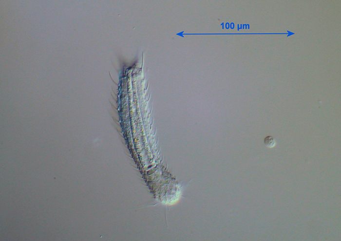
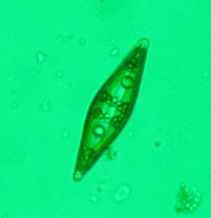 Large motile diatom ≥ 400 µm
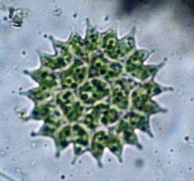 Pediastrum c. 200 µm
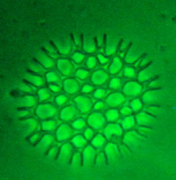 Pediastrum c. 200 µm
 Metopus high magnification 80–100 µm
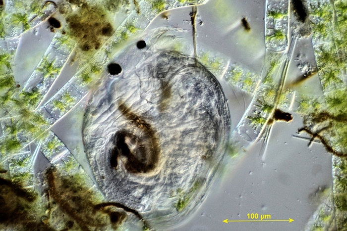
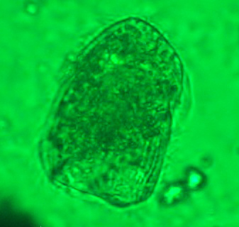
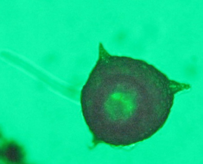 Testate amoeba c. 400 µl
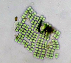 Raft of algal cells (Merismopedia)
Paul Smith’s specimens
Paul’s samples included what appeared to be a gill from a mayfly nymph, with various organisms attached to it.
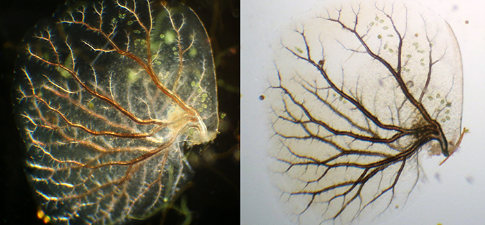 Gill of mayfly nymph (dark-ground and bright-field)
Acknowledgements
Our thanks to the Friends of Warnham Local Nature Reserve for allowing us to collect specimens and to use the Discovery Hub, and to Graham Matthews for organising the excursion.
Report and most photographs by Alan Wood, photomicrographs by Graham Matthews, Jacky McPherson, Mark Berry and Paul Smith.