

Arkwright Scholars Workshop
Saturday 23rd November 2019
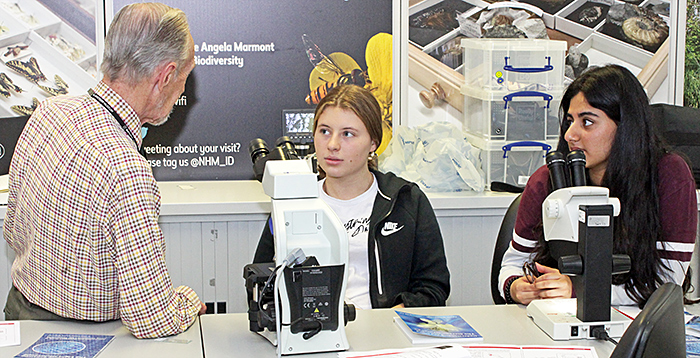
This was the sixth workshop for Arkwright Scholars on the uses of the optical microscope, held as before in the Angela Marmont Centre in the Natural History Museum in London. The Arkwright Engineering Scholarships are awarded by The Smallpeice Trust and support students through their A Levels or Scottish Highers and provide financial assistance to help them pursue engineering, computing or technical design at university or through a higher-level apprenticeship. The Scholarships are sponsored by industrial companies, universities, charitable trusts, trade associations, professional engineering institutions, the Armed Services, Worshipful Companies, industry regulators or personal donors. Quekett members Lisa Ashby, Pam Hamer, Chris Thomas, Dennis Fullwood, Paul Smith and Alan Wood assisted at the workshop. Tricia York and Tessa O’Shea from the Trust came to observe but couldn’t resist looking through the microscopes.
Introduction
Pam Hamer gave a brief introduction to vision, colour vision, light and lenses (including focal length, numerical aperture, resolution and magnification).
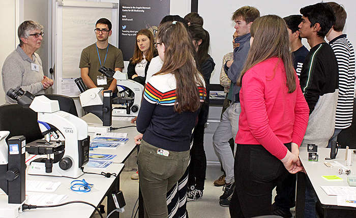 Pam Hamer’s introduction
She then showed how to use a lens (a linen tester that is easy to stand up) and a light to produce an image on a screen. Then how to use a second lens to magnify the image on the screen. She then removed the screen to show that the magnified image was clearer and brighter. This is the principle of a microscope, where the objective magnifies the object and produces an aerial image that is then further magnified by the eyepiece. The Scholars were then asked to do this themselves.
 Making microscopes
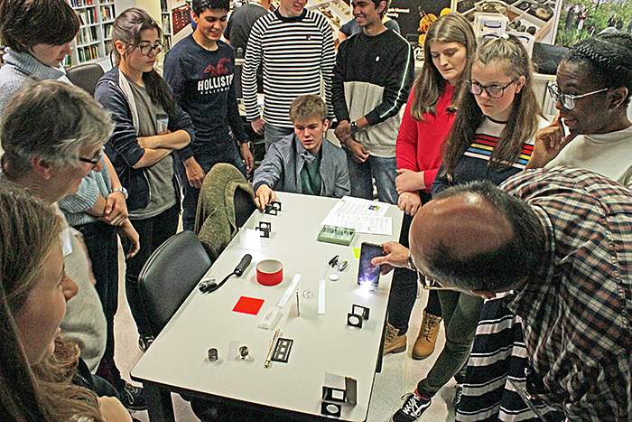
Pam also demonstrated fluorescence, where a substance absorbs light and then emits light of a longer wavelength. With the room darkened, Pam then shone a red laser through three diffraction gratings with different line spacings to demonstrate the direct transmission and first order diffraction.
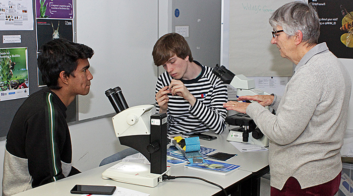
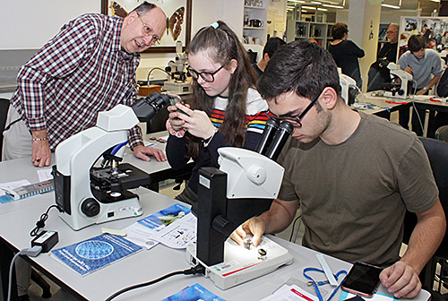
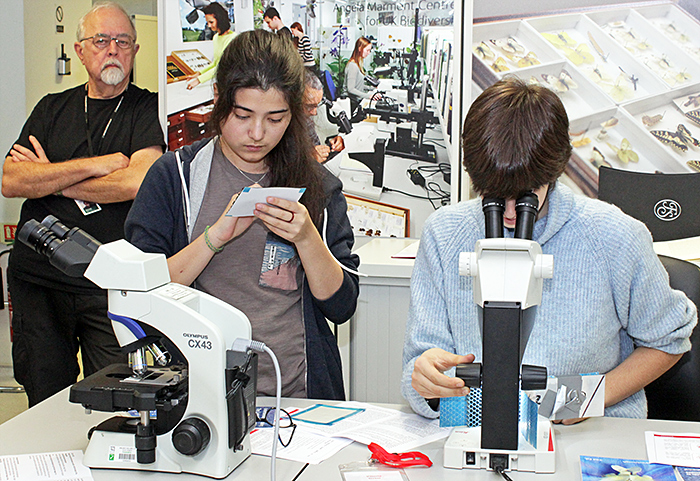
Pam explained the differences between compound microscopes and stereomicroscopes and their advantages and disadvantages, and explained how they should be adjusted to suit the user’s eyesight and to get the best results. Quekett members then helped the Scholars to set up the Leitz EZ4 stereomicroscopes and Olympus CX43 compound microscopes provided by the Museum.
 Lisa Ashby and Chris Thomas with Scholars
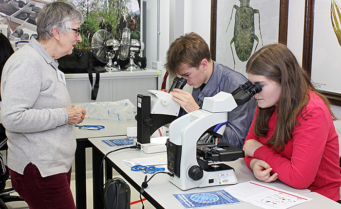 Pam Hamer with Scholars
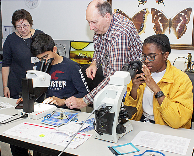
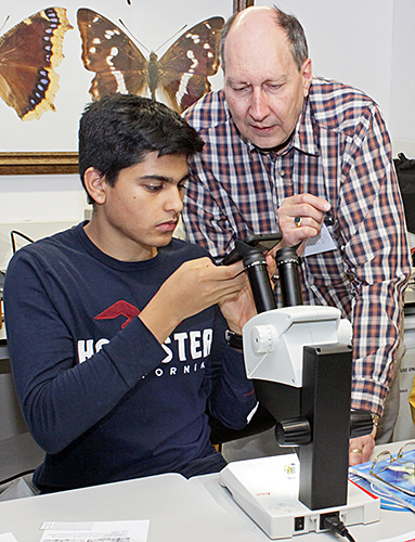
 Dennis Fullwood with Scholars
Materials science
Pam Hamer showed a PowerPoint presentation from Solvay (one of the sponsors of Arkwright) explaining the materials that they had supplied for the workshop.
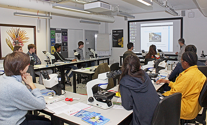 Solvay presentation
See Solvay’s presentation: click the arrow keys to move through the slides, click the bottom right icon for a larger version
Solvay had kindly provided some real materials for the Scholars to examine, including climbing drum peel test coupons (aluminium skin bonded to aluminium honeycomb with blue adhesive), carbon tooling laminate (layers of glass fibre fabric, carbon fibre fabric and resin) and samples of paint from the bonnet of a BMW car.
One skin of the climbing drum peel test coupons had been forcibly peeled off so that the blue glue could be examined to see whether there were any air bubbles, and to see the quality of the fillet (the shape of the adhesive where the skin and the honeycomb meet). The long working distance of the stereomicroscopes made them ideal for examining the coupons to see how well the glue had spread and whether there were any gaps in the glue.
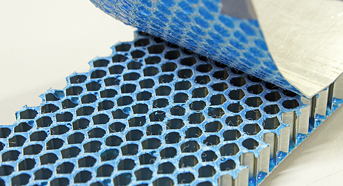 Climbing drum peel test coupon
One of the Solvay materials was a carbon tooling laminate containing carbon-fibre and glass-fibre mats; sections had been made from this, and samples of fibres were provided. The laminate was of a type used on high-performance boats and cars.
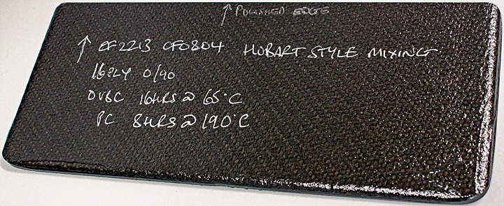 Carbon tooling laminate material
Sections of the laminate (approximately 22×6 mm) that had been mounted in resin and then polished were provided for examination under the Leitz stereomicroscopes.
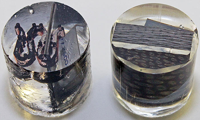 Paint section (left) and laminate section mounted in resin cylinders
There were visible defects in the laminate, including dry fibre (where the resin had not impregnated) and voids. With the aid of a piece of graph paper with 1 mm squares, the Scholars were asked to calculate the area of the field of view through a stereomicroscope and the area of the voids within the field of view, and then calculate the percentage of voids.
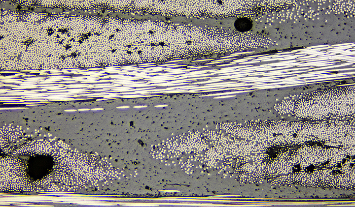 Laminate with voids (metallurgical microscope, vertical reflected light)
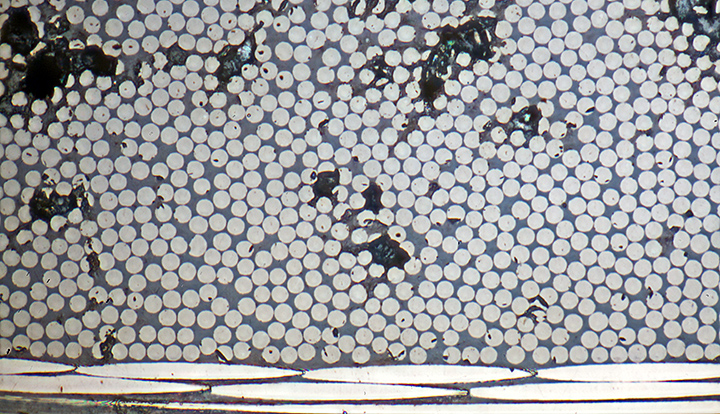 Sections through carbon fibres (7µm diameter) in laminate (metallurgical microscope, vertical reflected light)
Solvay also provided woven fabrics made of carbon fibre and glass fibre.
 Glass fibre and carbon fibre fabrics
The Scholars were provided with prepared microscope slides of carbon fibres and glass fibres (with human hair and spider silk for comparison), and were shown how to examine these using the Olympus CX43 compound microscopes. They used not only standard bright-field illumination but also 2 methods that provide enhanced contrast: dark ground and phase contrast. The Scholars were surprised to see the huge difference that lighting techniques can make to the appearance of a specimen under a microscope.
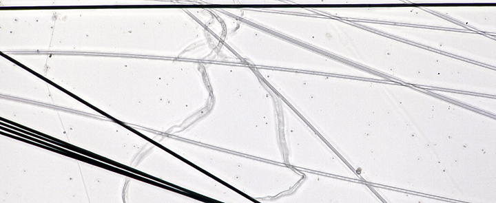 Carbon fibres, glass fibres and spider silk (compound microscope, bright-field illumination)
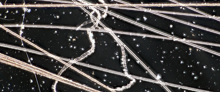 Carbon fibres, glass fibres and spider silk (compound microscope, dark-ground illumination)
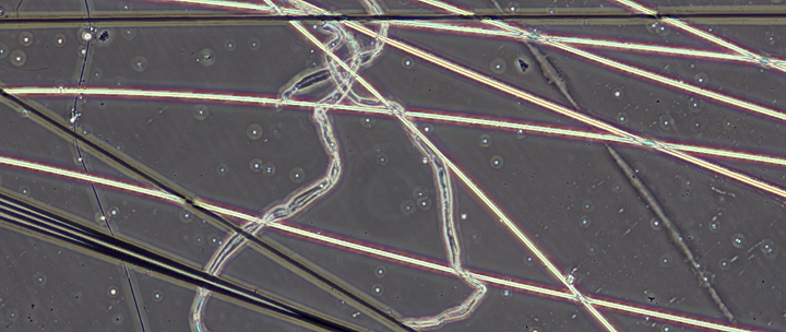 Carbon fibres, glass fibres and spider silk (compound microscope, phase-contrast illumination)
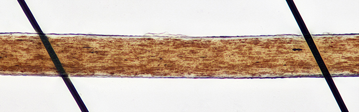 Carbon fibres (7 µm diameter) and a brown human hair (compound microscope, bright-field illumination)
Quekett members were on hand to show the Scholars how to adjust the microscopes to get the best views of the specimens.
 Pam Hamer with Scholars
 Chris Thomas with Scholars
 Paul Smith with Scholars
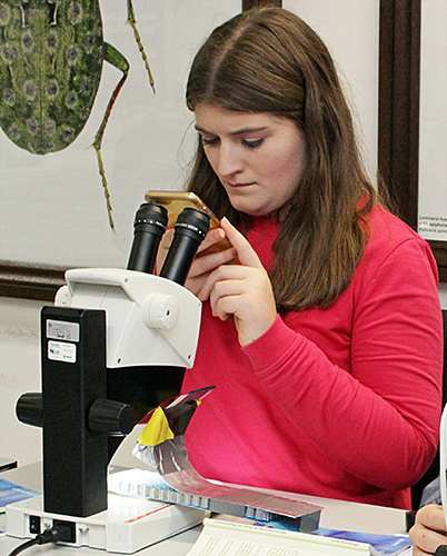
Some of the Scholars used their smartphones to record images through one of the microscope eyepieces; this is not easy, it needs a steady hand and you have to get the camera lens in exactly the right spot.
 Using a smartphone
 Using a smartphone
Making slides
Dennis Fullwood provided peacock corona feathers and locust legs for the Scholars to mount on microscope slides, with cutters, scissors, forceps, brushes, gum Arabic, PVA adhesive, labels and marker pens.
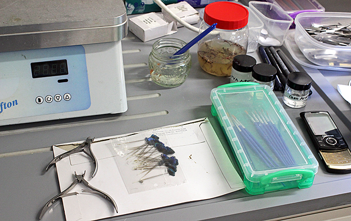 Materials for making slides
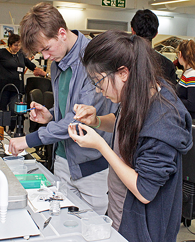 Making slides
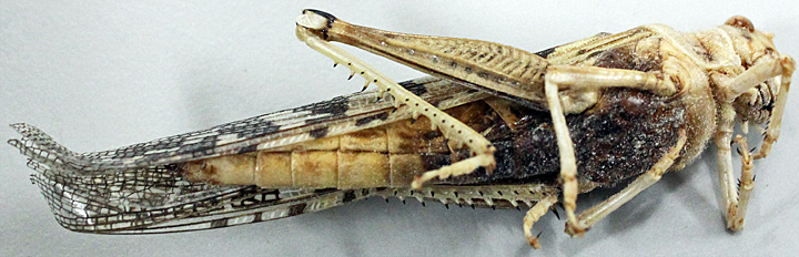 Locust
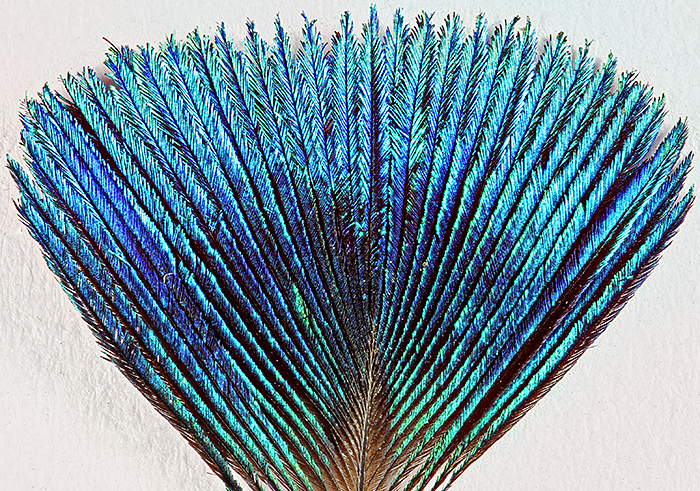 Peacock corona feather
Specimens, slides and microscopes
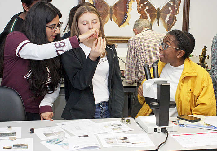
Some of the Quekett members brought microscopes and specimens to show to the Scholars.
 Scholars with microscope and specimens
Paul Smith brought a small digital microscope with a built-in screen (KKmoon Digital USB Microscope), and used it to show star sand.
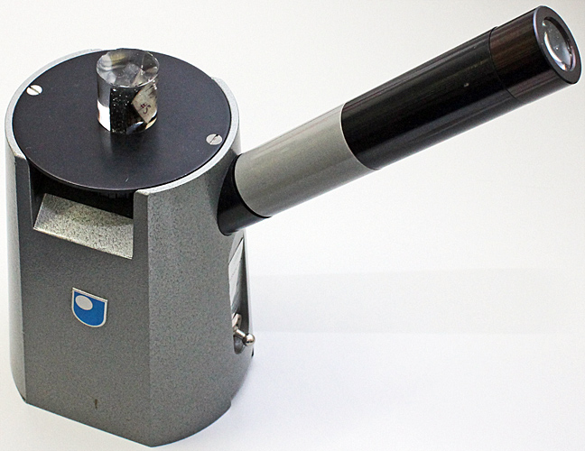
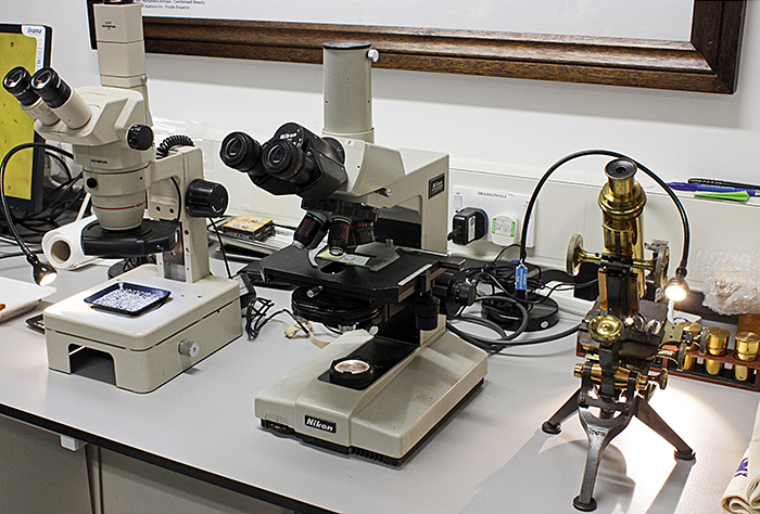
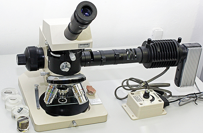
Pam Hamer brought a Gillett & Sibert Monolynx inverted metallurgical microscope. Biological microscopes such as the Olympus CX43 are designed to transmit light through transparent or translucent specimens, and the objectives are corrected for observing specimens through a 0.17 mm glass coverslip. Metallurgical microscopes use reflected light for observing opaque subjects and have prisms or semi-silvered mirrors to direct light through the objective onto a reflective subject and then back through the objective to the eyepieces. Their objectives are corrected for observing uncovered subjects.
Pam also brought lots of specimens, including a digital ‘chip’, car paint samples, cosmetics, coins, currency notes, rock samples with microfossils, cleaned microfossils, insects, and fossils.
 Pam Hamer’s metallurgical microscope and specimens
 Inverted Gillett & Sibert Monolynx reflected-light microscope, with laminate section viewed from underneath
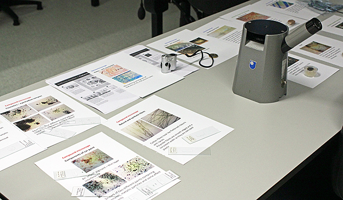
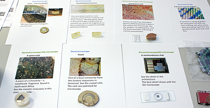 Some of Pam Hamer’s specimens and notes
Pam Hamer showed the Scholars where to look for micro writing on bi-metallic £1 coins and on the new plastic £10 bank notes.
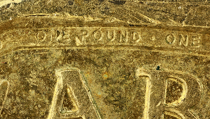 Micro writing on £1 coin (letters are 0.25 mm tall)
![]() Location of micro writing on £1 coin
Location of micro writing on £1 coin
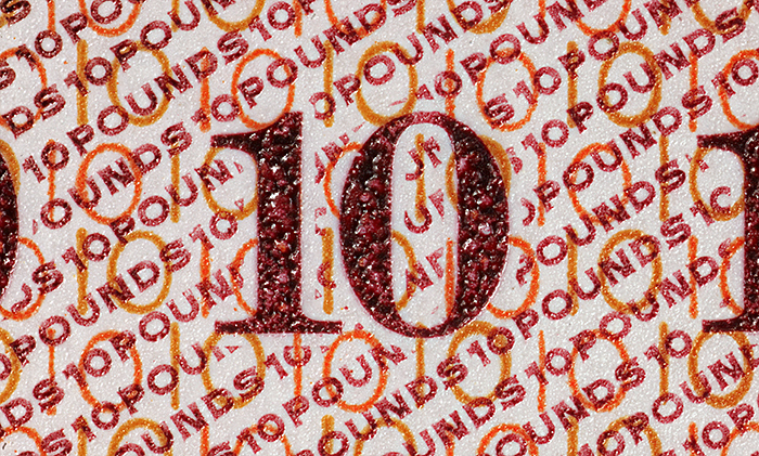 Micro writing on £10 note (letters are 0.25 mm tall)
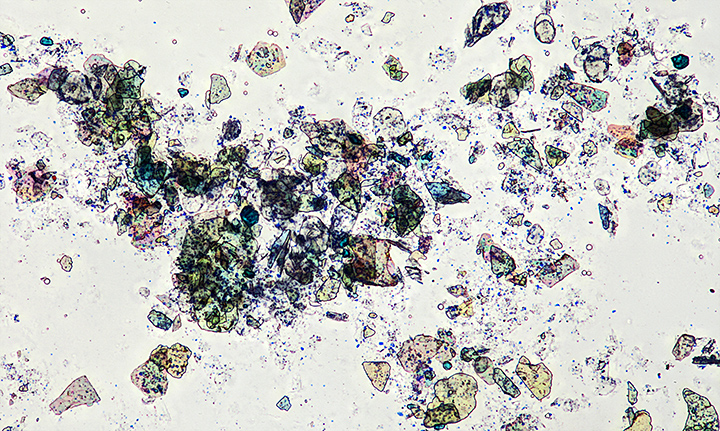 Eye shadow containing effect pigments
Dennis Fullwood brought his Olympus SZ4045 stereomicroscope, Nikon Labophot compound microscope, brass Watson Royal compound microscope and Chinese inspection camera, with insects and seeds in amber and slides from his collection and from the Quekett’s loan collection.
 Olympus SZ4045 stereomicroscope, Nikon Labophot compound microscope and brass Watson Royal compound microscope
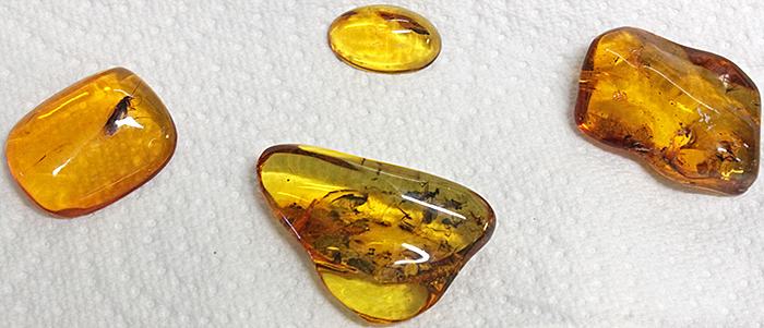 Insects in amber
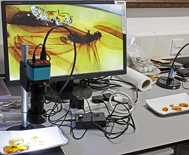 Insects in amber, imaged by a Chinese inspection camera and displayed on a monitor
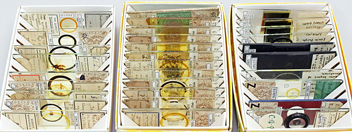 Slide sets
Alan Wood brought his Olympus BHMJ metallurgical microscope for viewing some metallurgical samples, the laminate sections, and some of Pam’s specimens.
 Olympus BHMJ metallurgical microscope
Two books by Quekett members were given to each Scholar: Understanding and Using the Stereomicroscope by Lewis Woolnough and Understanding and Using the Light Microscope by Chris Thomas and Lewis Woolnough.
Report and photographs by Alan Wood