Home Counties Meeting at Cobham
Saturday 24th February 2024
This was the second Home Counties Meeting held in Cobham and organised by Joan Bingley. It was held again in the main hall of Church Gate House Centre (where the South Thames Discussion Group met for several years) adjacent to St Andrew’s Church in Cobham, Surrey. The hall has a kitchen where we could help ourselves to tea, coffee and biscuits. We were pleased to welcome members of the Elmbridge Natural History Society and the Holmesdale Natural History Club that Joan had invited to attend. The day started with refreshments, followed by two talks, a break for lunch, and an afternoon of exhibits and gossip.
Talks
Bevil Templeton-Smith: “Art photography using a microscope”
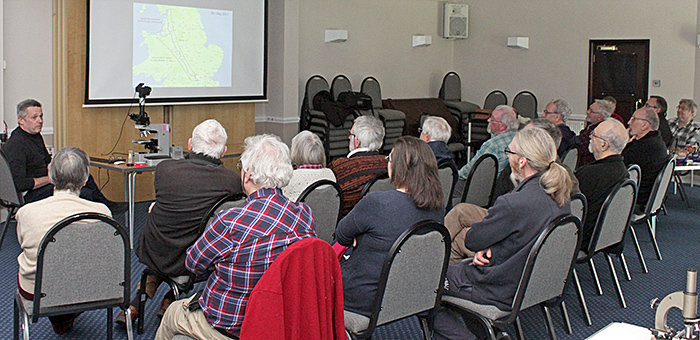 Audience for the talks
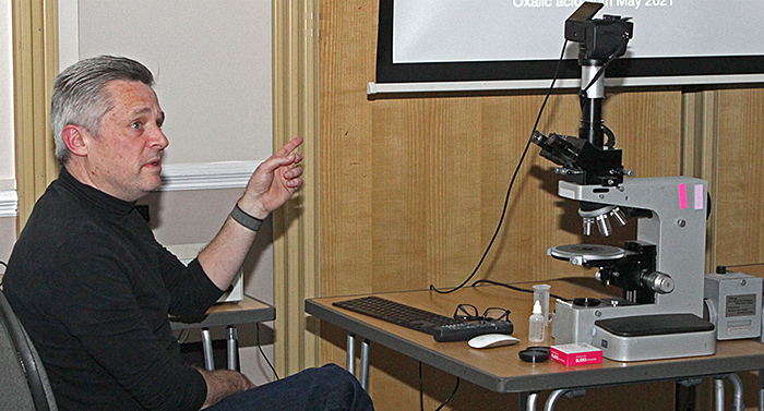 Bevil Templeton-Smith
Bevil has been a keen photographer for many years, with interests including astrophotography and macrophotography, but did not consider using a microscope until he saw a colourful image of an iridescent seed of purslane (Portulaca oleracea) taken by Yanping Wang using a stereomicroscope.
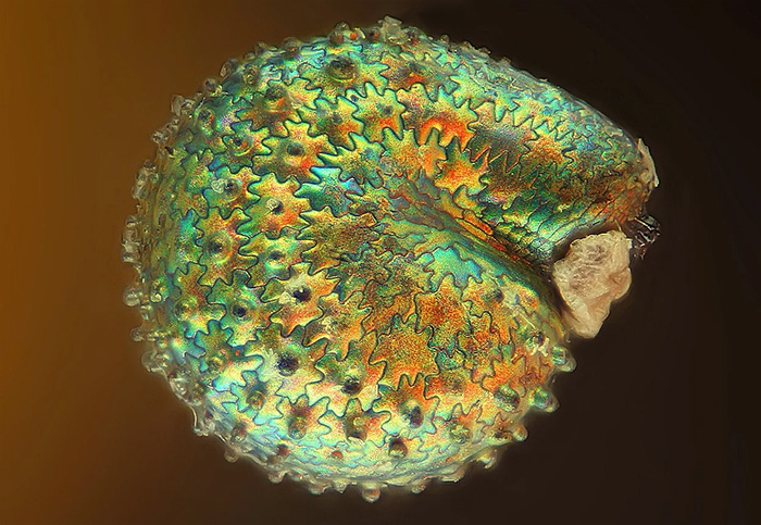 Purslane seed [By Yanping Wang]
Inspired by this image, he bought a Baker stereomicroscope on a boom stand and a packet of purslane seeds, but his photos showed only a dull brown seed. He did not give up, he bought his first trinocular Leitz Orthoplan in 2021, made an adapter for his Sony α7R IV mirrorless camera, and started taking photographs of crystals. It took him a while to realise that he needed a suitable eyepiece to get edge-to-edge sharpness, even with stacked images, and he obtained one from another Leitz that came with an Orthomat camera. He extracted the Vario-Orthomat zoom photo eyepiece that fits the 38 mm photoport and constructed an adapter to place his camera at the correct location. Then there was no stopping him, and he bought a succession of Leitz microscopes to obtain the objectives, polarisers, analysers and stages that he needed. For retarders, he uses mica sheets of various thicknesses and an assortment of plastic films. He examines each slide closely, and often selects just a tiny portion to photograph. He experimented with lots of chemicals, and his photographs started to attract attention. In 2023, there was an exhibition of his work called “Polychromo” at Alveston Fine Arts gallery in Notting Hill.
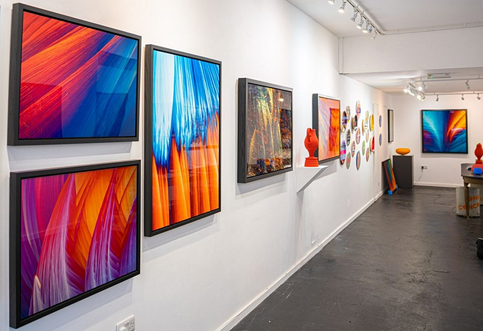 “Polychromo” at Alveston Fine Arts
Also in 2023, Bevil was awarded non professional Fine Art Photographer of the Year in the International Photography Awards. He still wants to take a good photograph of a purslane seed, and now he knows that he should probably use a fresh seed, not a dried one.
You can see several of Bevil’s crystal photomicrographs on his Facebook and Instagram pages.
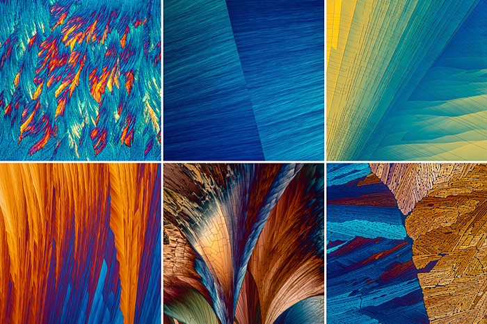 Some of Bevil’s crystal photomicrographs on Instagram
Robert Ratford: “Stereoscopic microscopes”
Robert gave a short talk on stereomicroscopes, which first became available in 1896. Since then, their field of view has increased, the range of magnifications has increased, transmitted light has become easily available, and zoom objectives have been introduced. They provide a much greater working distance and depth of field than compound microscopes, but with lower resolution and lower maximum magnification.
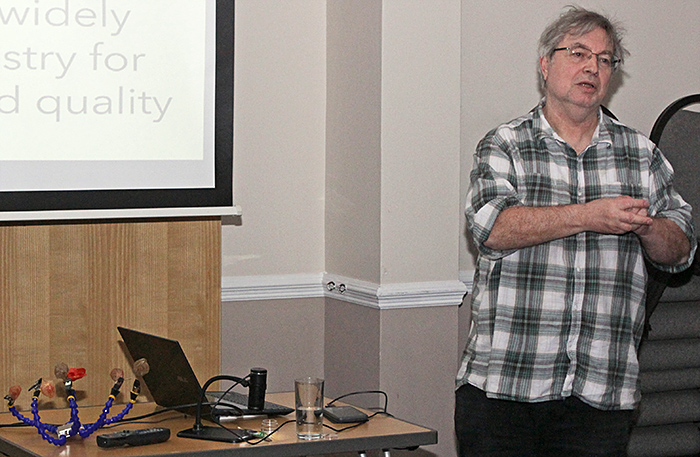 Robert Ratford
Lunch
For lunch, members could either bring their own or opt for fish and chips from a local shop, kindly ordered and collected by Joan Bingley.
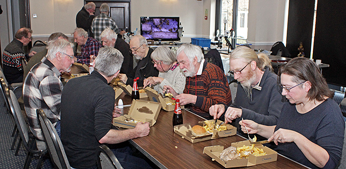 Lunch (fish and chips)
Gossip
As with any Quekett meeting, there was gossip before the talks and during lunch, as well as in the afternoon.
Lisa and Nigel Ashby brought two Watson items that they had recently acquired. One was a “School of Mines” Petrological Microscope, with a photocopy of a catalogue entry and some modern slides. The smooth brown surface of the stage was interesting, it could be Bakelite.
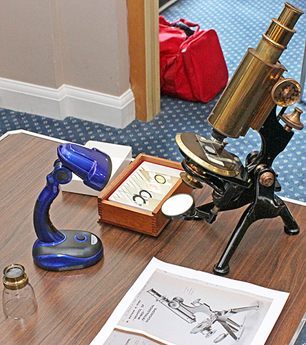 Watson “School of Mines” Petrological Microscope
The second item was the Electric Heating Stage, in its original felt-lined box complete with thermometer and type-written instructions. It would need a pump and a power supply before it could be tried.
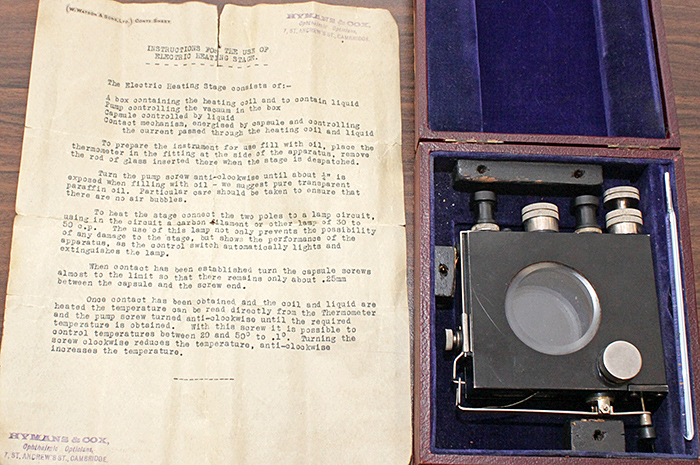 Watson Electric Heating Stage
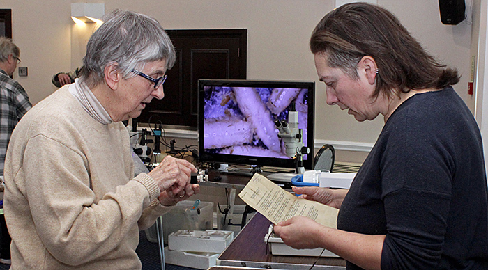 Pam Hamer and Lisa Ashby (right)
Mark Berry likes assembling unusual components into usable equipment and brought some examples.
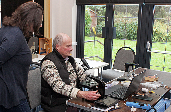 Lisa Ashby and Mark Berry
Mark found a bare Nikon stand, and added a Vickers binocular head, a pair of Bausch & Lomb eyepieces, a large Leitz or Zeiss stage (for which he machined a missing part), and finally some Nikon short-barrel objectives.
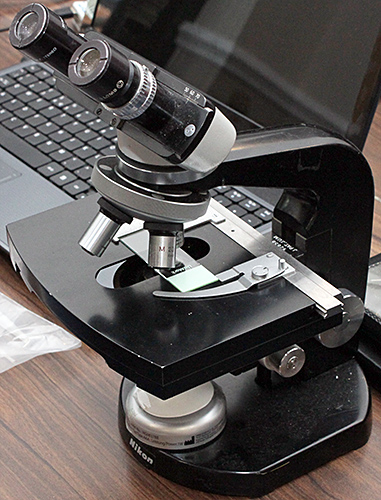 Nikon microscope
Mark also brought an unusual Myacope zoom monocular microscope to which he had fitted an adapter for a smartphone.
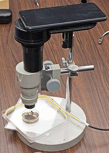 Myacope microscope with LED side illumination.
Mark has recently started looking at the inhabitants of moss, and showed his first attempt at lighting water in a small Petri dish from the side. It uses a strip of LEDs mounted inside a square Petri dish
Mark also showed how he had made an adapter to hold a small camera a little way above a microscope eyepiece. It uses a metal tube and parts of bicycle inner tubes.
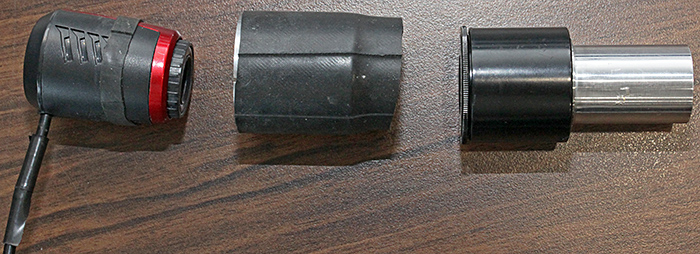 Camera attachment
Joan Bingley brought her small Brunel stereomicroscope so that we could examine some feathers and some slides of feathers.
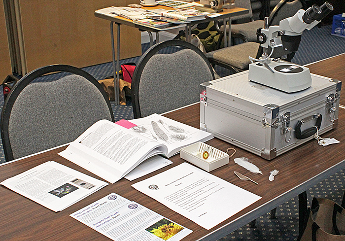 Joan Bingley’s exhibit
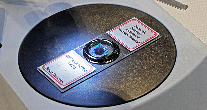 Peacock feather mounted by Brian Darnton
Joan also brought a book that explains the details of the structure of feathers:
- Manual of Ornithology: Avian Structure and Function by Noble S. Proctor & Patrick J. Lynch
Stephen Durr brought some material from Gordon Brown’s well-established compost heap and shook it through a sieve to extract pseudoscorpions.
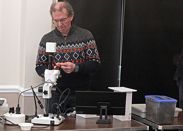 Stephen Durr with his exhibit
For observing the pseudoscorpions, Stephen brought a trinocular stereomicroscope that had 2 flexible arms with LEDs for reflected light and also LED transmitted light. The microscope was fitted with a Tucsen TrueChrome Metrics camera.
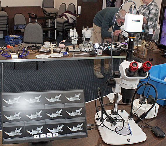 Stephen Durr’s exhibit
Phil Greaves brought his beautiful black trinocular Leitz Ortholux equipped with the Smith T (transmitted) differential interference contrast objectives and condenser. This system produces excellent images but was difficult to manufacture because the second prisms had to be fitted inside the objectives at the rear focal plane. The objectives also had to be precisely orientated and fitted in the nosepiece at the factory, and only 25×, 40× and 100× objectives were produced.
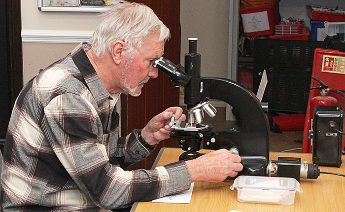 Nigel Williams with Phil Greaves’ exhibit
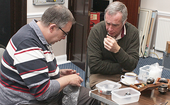 Jonathan Crowther and Phil Greaves (right)
Pam Hamer brought two Vickers polarising microscopes, one equipped for transmitted light (for slides of thin rock sections) and the other for reflected light (for polished rock specimens). To demonstrate transmitted light, Pam used a commercial slide of basalt that included olivine, pyroxene and plagioclase crystals of felspar. To demonstrate reflected light, Pam used a polished section of a coal ball that had been prepared by the late Jamie Nelson.
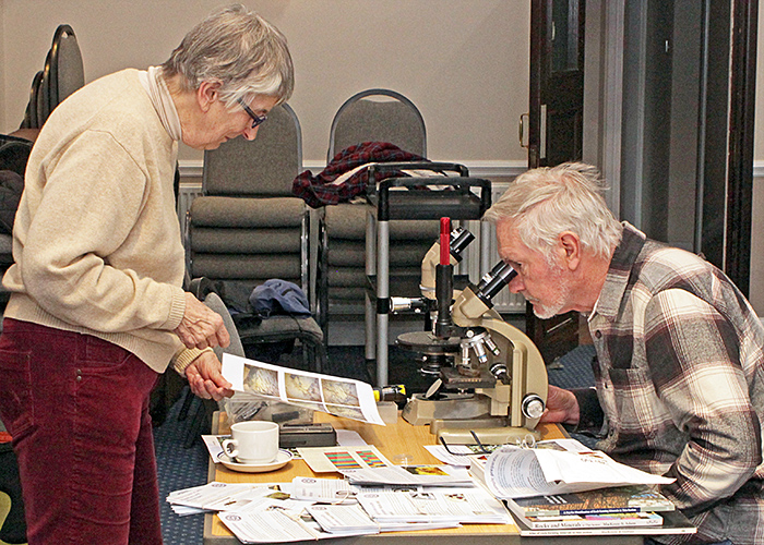 Pam Hamer and Nigel Williams
It is not easy to identify minerals in geological thin sections, and Pam showed four books that she uses, including an identification key:
- A Key for Identification of Rock-Forming Minerals in Thin Section by Andrew J. Barker
- Atlas of the Rock-Forming Minerals in Thin Section by W. S. Mackenzie & C. Guilford
- Rocks and Minerals in Thin Section: A Colour Atlas by W. S. MacKenzie, A. E. Adams & K. H. Brodie
- Rocks & Minerals: The Definitive Guide by DK
Graham Matthews brought a box of slides that he has made of specimens found in leaf litter at Warnham Local Nature Reserve. He provided a Biolux school microscope (modified by adding LED illumination and a mechanical stage) so that we could examine the slides.
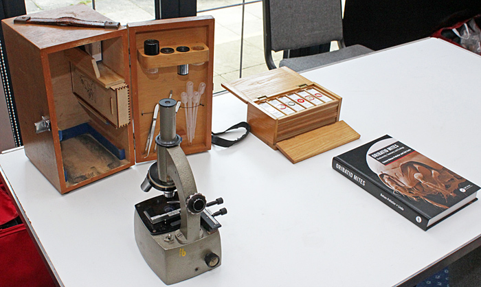 Graham Matthews’ exhibit
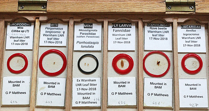 Some of Graham Matthews’ slides
Mites are some of the most difficult specimens to identify, and Graham showed a book that he has bought to help identify Oribatid mites to at least genera.
- Oribatid Mites: Biodiversity, Taxonomy and Ecology by Valerie Behan-Pelletier & Zoë Lindo
Robert Ratford filled two tables with his exhibit. One table was dominated by a large television showing live images of the maggots that are used as bait by anglers. Robert brought four stereomicroscopes, one by Carl Zeiss Jena and three modern Chinese ones, two of which were trinocular. He also brought an assortment of cameras, digital microscopes and LED lighting, and a gadget that can support six specimens on its flexible arms. Robert’s specimens included feathers and whole bird wings.
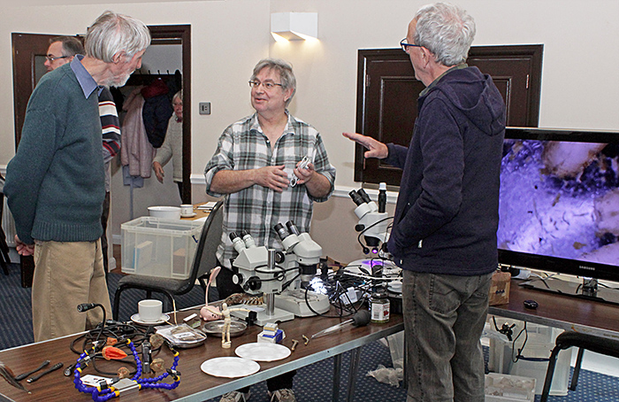 Robert Ratford with Brian Spooner (left) and Martin Parnham (right)
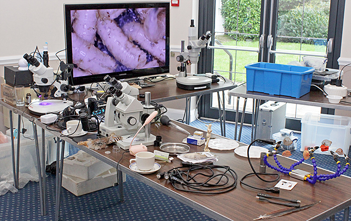 Robert Ratford’s exhibit
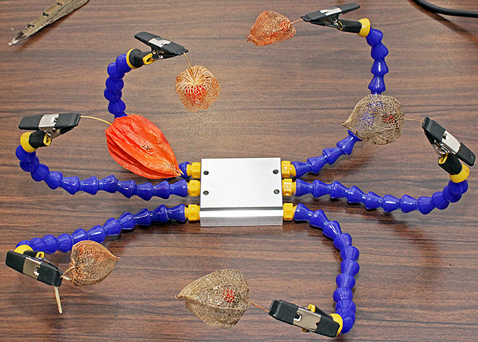 Specimen holder
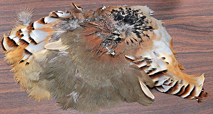 Bird wing
Bevil Templeton-Smith brought one of his Leitz Orthoplans and his Sony α7R IV mirrorless camera and used them for live demonstrations during his talk in the morning. In the afternoon, he was kept busy answering more questions about his equipment, his photomicrographs of crystals, his techniques, and the software that he uses (Helicon Focus, Adobe Lightroom and Adobe Photoshop). You can see several of his crystal photomicrographs on Instagram.
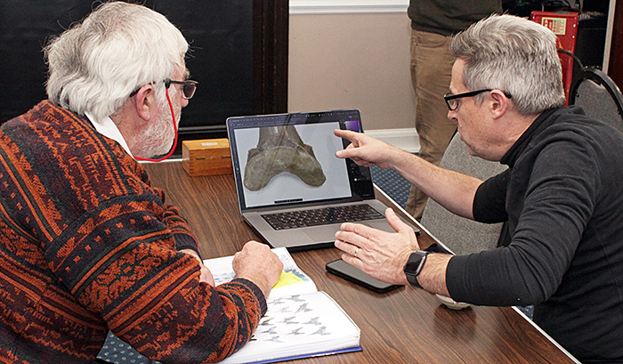 Trevor Batchelor (left) and Bevil Templeton-Smith
Acknowledgements
Our thanks to Joan Bingley for organising the event, providing the refreshments and collecting the fish and chips, and to all those who got out and put away the tables and chairs.
Report and most photographs by Alan Wood

