

Wimbledon Common Nature Club
Sunday 8th January 2023
The Wimbledon Common Nature Club is run by Auriel Glanville and two teenage assistants (Alexander and Oliver Mallett) and welcomes children from 6–14 years old to come and discover the world of nature on the Common. They meet for 2 hours each month in the Information Centre, the same venue as used by Quekett members on excursions, the Weekend of Nature and the Open Day.
As part of the Quekett’s outreach programme, Paul Smith and Alan Wood took an assortment of microscopes and some interesting slides and specimens to show the children. As usual, some of the parents were keen to have a look too.
Alan took his Olympus SZ4045 stereomicroscope with an LED ring-light, a small stereomicroscope (the STX Stereo Microscope sold on Amazon by GT Vision) and a Natural History Museum Microscope. He brought some lichen, jay and parakeet feathers, a butterfly wing and several microscope slides, and collected bits of brambles and stinging nettles from the Common.
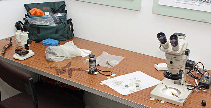 Alan’s microscopes
Paul took his Swift FM-31 portable microscope, a Telmu inverted microscope and a digital microscope with a built-in screen. Paul’s specimens included an insect in a polished piece of amber, insects embedded in resin blocks and a watch. He used the FM-31 to demonstrate how polarising filters and plastic sheets can add colours to some subjects (see the last image on this page).
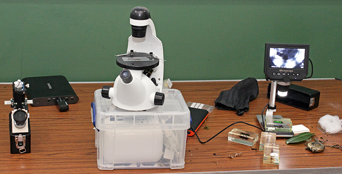 Paul’s microscopes
When everyone had arrived, Auriel led us on a short walk along the muddy paths through the woods near the Centre. The children collected lots of items to take back and examine under the microscopes, including rotten wood, bark, toadstools, dead fern fronds, mosses and lichens
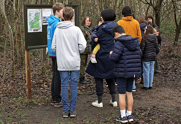 Start of the walk
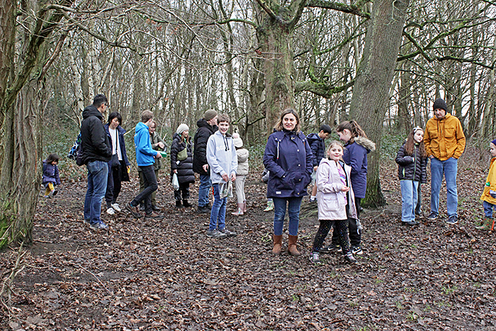 Walking in the woods
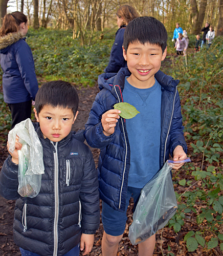 Collecting in the woods [by Auriel Glanville]
We got back to the Centre just as it was starting to rain, and started looking at the specimens.
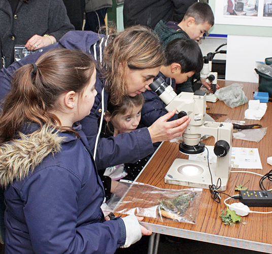 Family with microscopes
 Zooming in for a closer look [by Auriel Glanville]
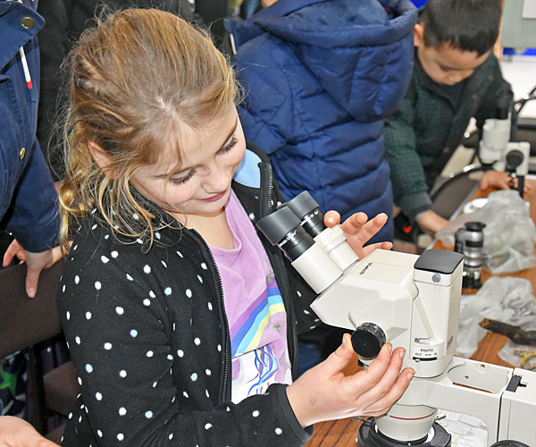
 Two girls with microscopes [by Auriel Glanville]
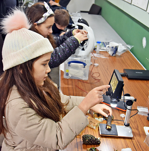
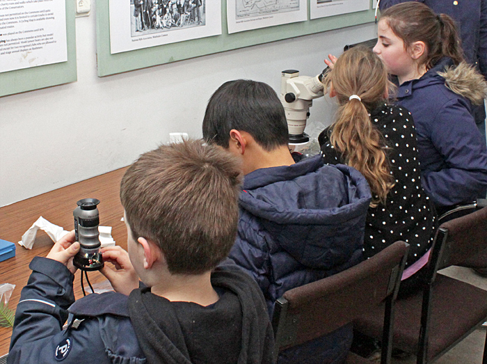 Four children with microscopes
One of the parents kindly took a group photograph for us:
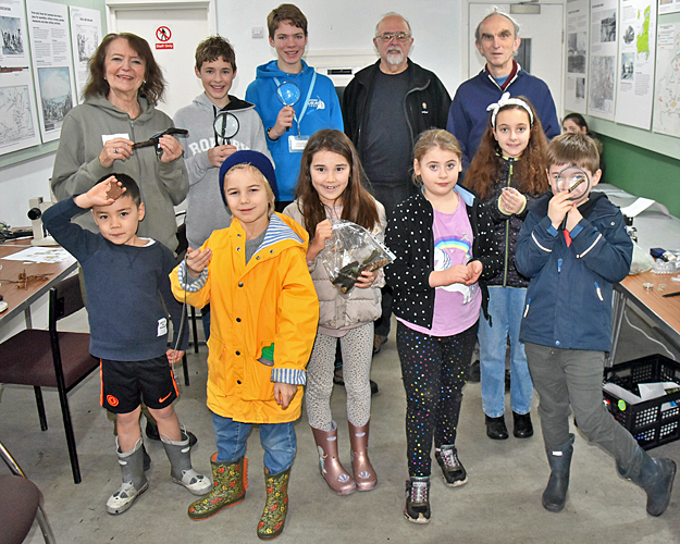 Group photograph
Back row: Auriel, Oliver, Alexander, Paul and Alan
Here are photographs of some of the specimens:
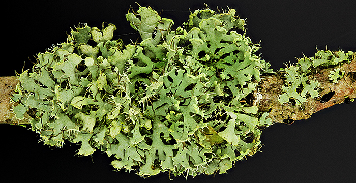 Green lichen on an oak twig
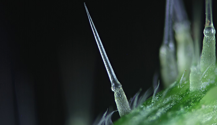 Hairs on stem of a stinging nettle
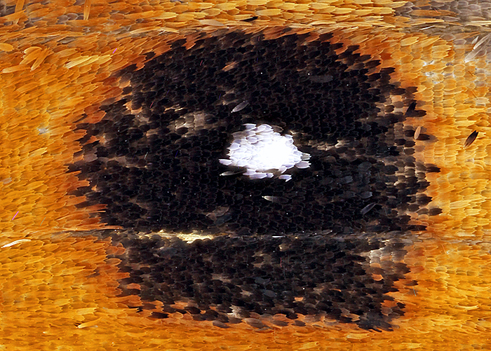 Eye-spot of a small heath butterfly (1.2 × 1 mm)
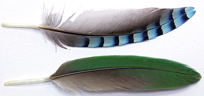 Feathers of a jay and a ring-necked parakeet
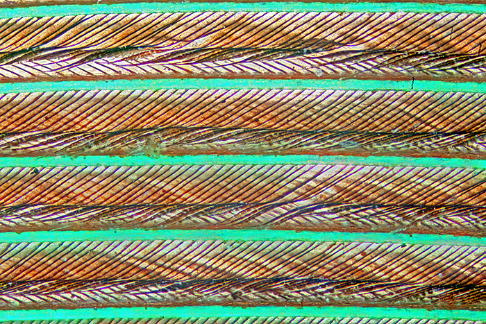 Feather of a ring-necked parakeet
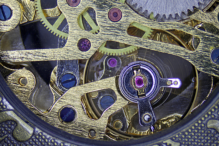 Watch movement
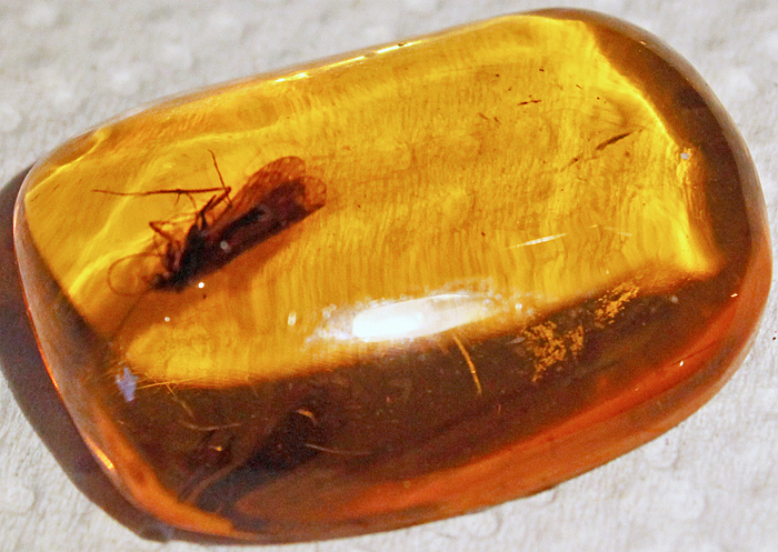 Insect in Baltic amber, about 40 million years old
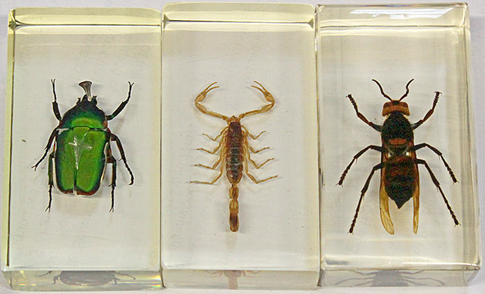 Insects and a scorpion in resin
The specimens on microscope slides need to be very thin. Small ones, like a flea, a flatworm, a foraminiferan or a hydra, can be mounted whole. Larger specimens need to be sliced into thin sections, like an earthworm, a rabbit embryo, a snail or the root or stem of a plant. The sections are so thin that they are almost colourless, so they are usually stained to make them easier to see.
Here are some of the microscope slides:
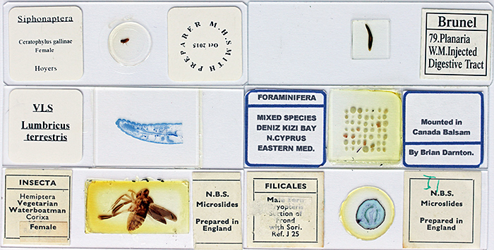 Microscope slides
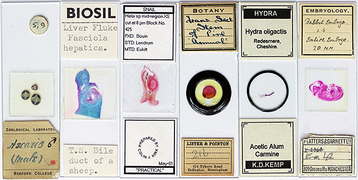 Microscope slides
And here are some of the specimens on the microscope slides, photographed through a microscope:
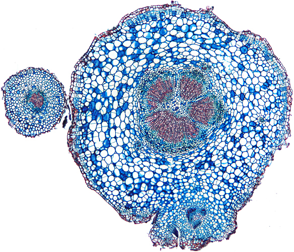 Stained cross-section of the root of a daisy
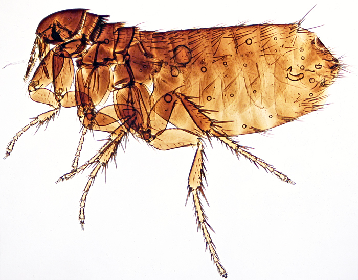 European chicken flea (Ceratophyllus gallinae)
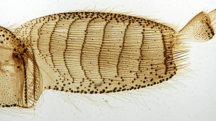 Pollen basket on third leg of a worker honeybee
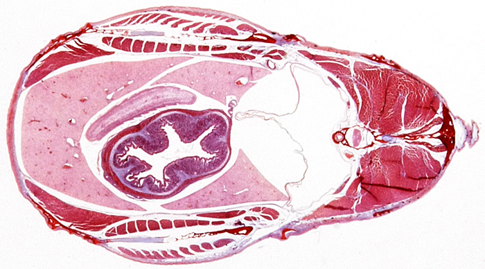 Stained cross-section of a tapeworm in a stickleback
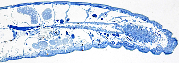 Stained longitudinal section of an earthworm (Lumbricus terrestris)
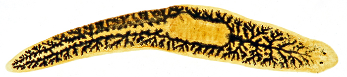 Planarian (flatworm), with black dye injected into its gastrovascular cavity

Stained longitudinal section of a rabbit embryo
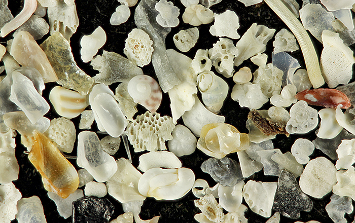 Shell sand from Dog’s Bay on the west coast of Ireland
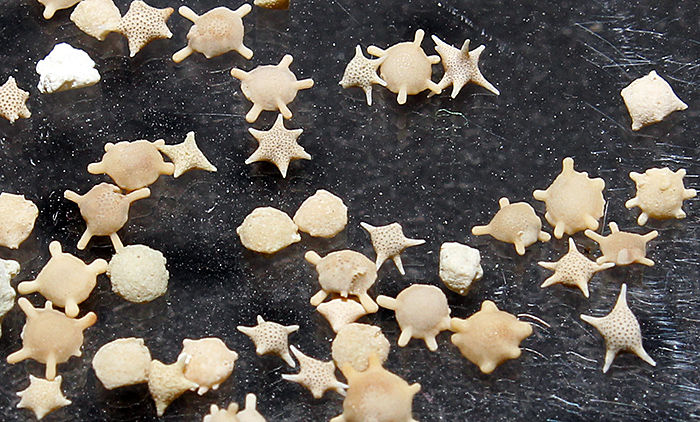 Star sand from Okinawa, Japan
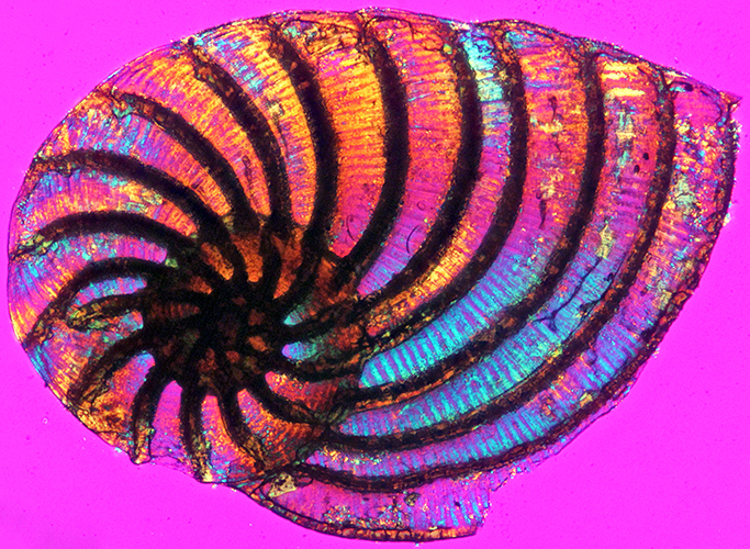 Foraminifera from North Cyprus (length 1.35 mm, colours produced by crossed polarising filters and a plastic sheet)
Report and most photographs by Alan Wood