

Annual Exhibition of Microscopy
Saturday 14th October 2023
The main report is available below. Too keep it to a manageable size, you can see all of the photomicrographs, videos, slides and artwork that were submitted for awards on these separate pages:
- Barnard Awards (Artistic)
- Barnard Awards (Technical)
- Barnard Awards (Videos)
- Eric Marson Awards
- Artwork Awards
- Celebrating our members – David Linstead
Quekett members: There is a value-added version of this report in the password-protected Members’ area of the website. This provides larger versions of the Barnard and Marson photomicrographs, includes the comments made by the judges, and lets you watch the videos of the lectures.
The Club’s Annual Exhibition of Microscopy (Quekex), was held for the third time at Elm Court Youth and Community Centre, Potters Bar, Hertfordshire. As usual, there were demonstrations and exhibits by members, displays of the photomicrographs, videos, slides and artwork submitted for awards, lectures, and plenty of gossip.
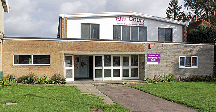 Elm Court Youth and Community Centre
In accordance with tradition, Club President Terry Hope opened the meeting by sounding the Quekett horn.
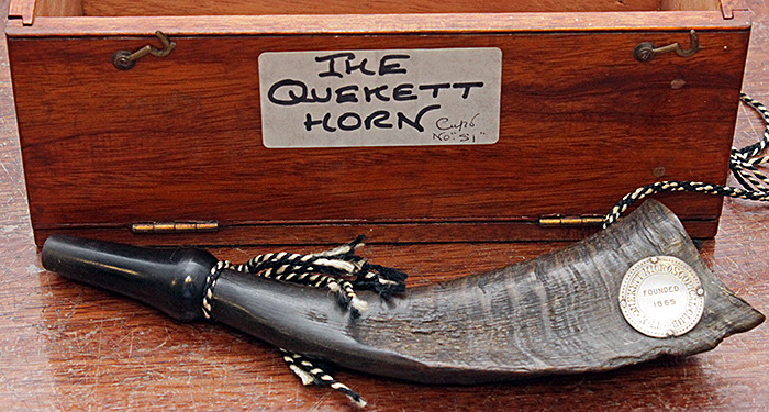 The Quekett horn
Exhibits
Lisa and Nigel Ashby brought a black and brass Watson Edinburgh microscope that was made with a special stage for E. M. Nelson, shown with a contemporary lamp and stand.
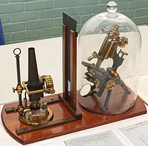 Watson Edinburgh made for E. M. Nelson
They also showed several other Watson microscopes, including two Bactils of different generations with old and new methods of displaying images to an audience.
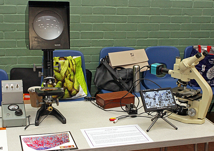 Displaying images from Watson Bactil microscopes
Nigel also showed a dry mount slide of sand from Dornoch Beach in Scotland, the first slide that he has made.
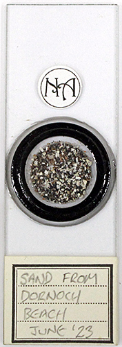 Dry mount of sand from Dornoch Beach
Joan Bingley brought some slides of Sphagnum that we could examine using a small stereomicroscope, some books and some old microscopes.
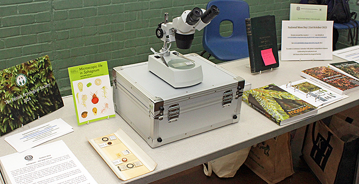 Joan Bingley’s exhibit
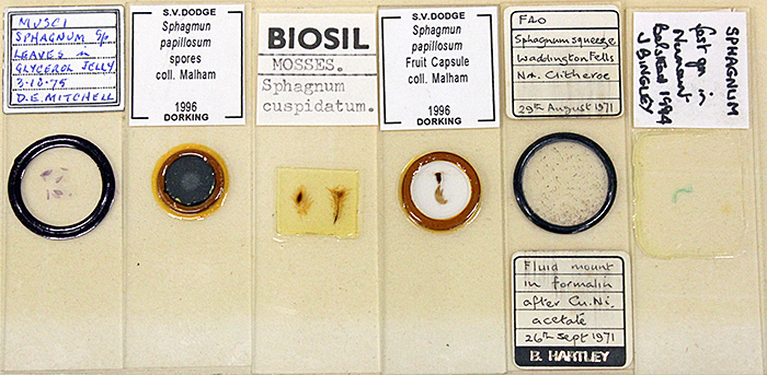 Slides of Sphagnum
With her notes and slides, Joan showed a relevant book, Microscopic life in Sphagnum by Marjorie Hingley.
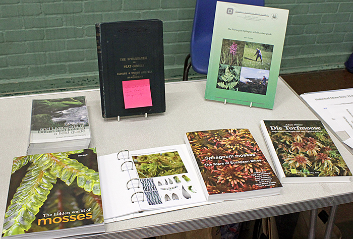 Joan Bingley’s books on mosses
The other books that Joan displayed included The Hidden World of Mosses by Neil Bell, Sphagnum mosses – The Stars of European Mires by Jukka Laine et al., Die Torfmoose: Südwestdeutschlands und der Nachbargebiete by Adam Hölzer, The Norwegian Sphagna: a field colour guide by Kjell I. Flatberg (free PDF), Mosses and Liverworts of Britain and Ireland: a field guide by Ian Atherton, Samuel D. S. Bosanquet & Mark Lawley and The Sphagnaceae or peat-mosses of Europe & North America by R. Braithwaite.
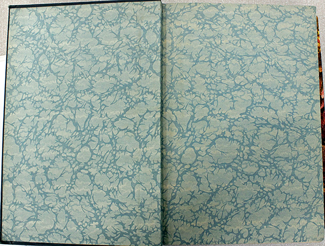 Endpapers of Braithwaite, based on the hyaline cells in a Sphagnum leaf
Joan also showed three brass and glass microscopes, a Watson stereo from 1932, a Koristka compound from around 1920, and a Zeiss jug-handle compound from the 1920s, all with their wooden boxes.
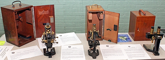 Joan Bingley’s brass and glass microscopes
Gordon Brown demonstrated how quick and easy it is to make slides using LOCA as the mountant with a UV lamp to set it.
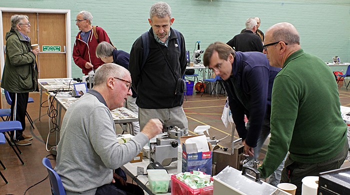 Gordon Brown (seated) with his exhibit
Gordon brought several rotary, sledge and rocking microtomes, including a Cambridge Frigistor (complete with its control box) that is used to cut sections of frozen specimens.
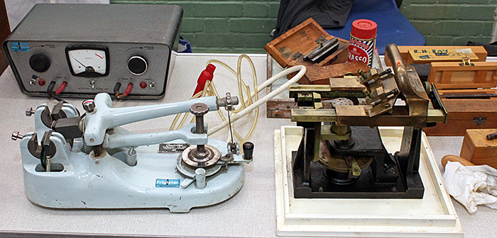 Microtomes
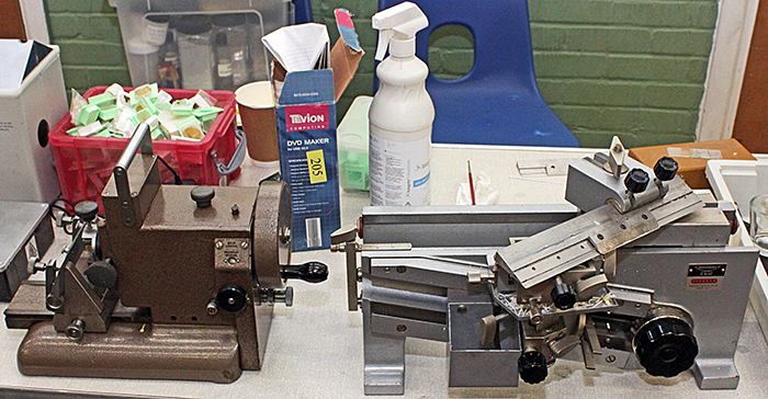 Microtomes
He also showed two Shandon Elliott sharpeners for microtome knives that he has worked out how to use. Gordon would like to hear from anyone who has experience using these machines.
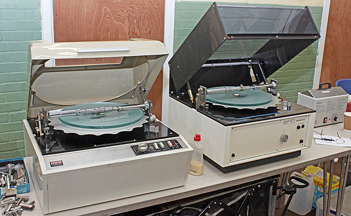 Microtome knife sharpeners
Douglas Downer-Smith has some thin section slides of a meteorite, rocks and minerals, including ones bought from Trevor Emmett at Microscopium, and wanted to be able to examine the whole specimens by transmitted light using crossed polarisers. He uses his AmScope stereomicroscope (a “simul-focal” model that allows the photo port to be used at the same time as both eyepieces) with a 0.75× supplementary objective to increase the field of view. Illumination is provided by a Rybozen slide scanner with its 4″×5″ LED panel masked down to 75×75 mm with black card. A Hoyarex 611 linear polariser sits on the panel to provide a polariser. The rotatable analyser is a normal linear polariser for a camera, with a 49 mm thread and a step-up adapter so that it fits the 48 mm thread on the objective.
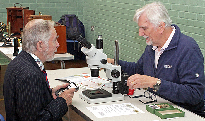 Terry Hope and Douglas Downer-Smith (right)
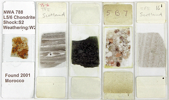 Douglas Downer-Smith’s slides
Douglas also provided notes and a book explaining igneous, sedimentary and metamorphic rocks.
For identifying diatoms, Douglas likes the key in A guide to the morphology of the diatom frustule by Horace Barber & Elizabeth Haworth. To suit his way of working, he is transcribing the booklet into a pictorial flow chart, and he showed the first draft.
David Furness is one of the few Quekett members with an electron microscope, the subject of his lecture at Quekex in 2021. This time, he brought two trinocular Zeiss Standard microscopes, one equipped for phase contrast and the other for differential interference contrast (DIC). The one for phase contrast was fitted with an HD Pro Webcam C920 from which David had removed the lens, mounted above a 20× eyepiece and sending images via USB to a laptop. The one for DIC was fitted with a modern eyepiece camera (with a 0.5× reducing lens), sending images via USB to ArcSoft Webcam Companion on a laptop.
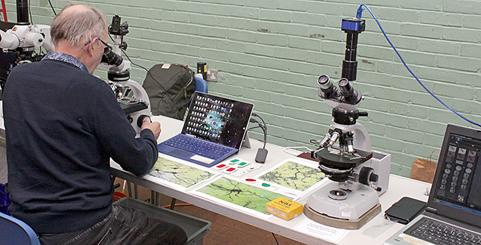 David Furness with his exhibit
David also brought some of his anaglyph 3D stereo photographs of nerves, with red/green glasses for viewing them. He uses the free Picolay program to take a stack of images and then generate the pair of images for viewing in 3D.
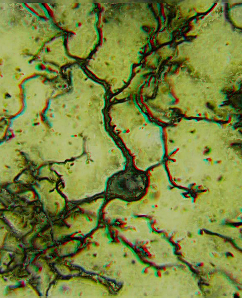 3D stacked image of nerves [By David Furness]
Click the image for a larger version. Use red/green anaglyph 3D glasses to view this image (red on right eye).
Steve Gill brought several pairs of coverslip polishers, one pair of slide polishers and some real and synthetic chamois leather. Flatters & Garnett used to sell coverslip polishers, and Colin Kirk devised a way of making them from the front panels and knobs of old drawers, using a 2½″ hole cutter, a small file and some chamois leather. Steve brought a genuine Flatters & Garnett pair; he made all of the others using Colin’s method.
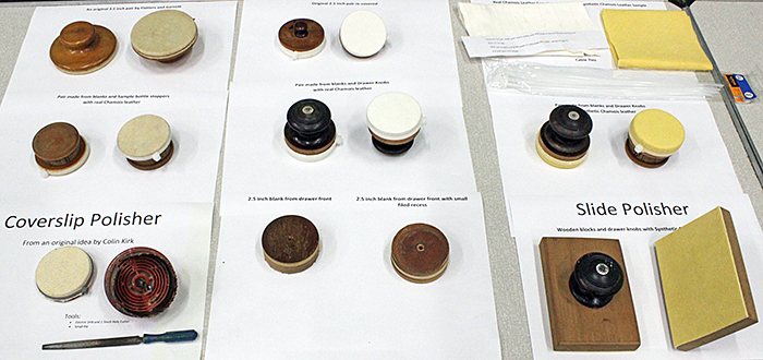 Polishers for coverslips and slides
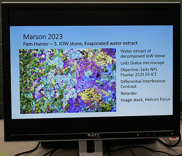
Pam Hamer collected mudstone in March 2022 from Compton Bay in the Isle of Wight. She spotted a stone with distinctive layer structure indicating a sedimentary rock origin, but she could see some shiny crystals so she took it home to examine it, and she has been observing and recording its gradual decomposition.
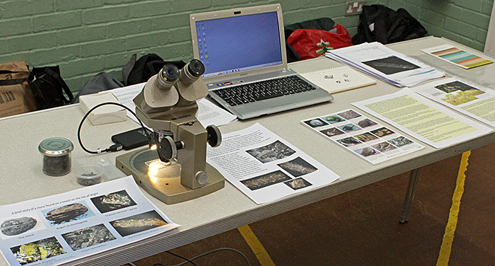 Pam Hamer’s exhibit
 Pam Hamer’s microscope
This PowerPoint presentation documents Pam’s observations from March 2022 to October 2023:
Click the arrows to move through the slides. Click the symbol at bottom right for a larger version.
By the time of the Cobham meeting in March 2023, the original mudstone had degraded to plant material, pyrite and ferrous sulfate. Since then, the stone has continued to disintegrate leaving a powdery grey residue. The remnants included dark, hard and shiny fragments that resembled stem residues. Pam believes they are likely to be the pyritised fossils of plants.
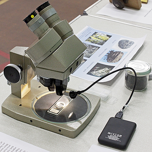
All that glitters… An interesting rock from a former copper mine in Parys Mountain in Anglesey, a fascinating geological site.
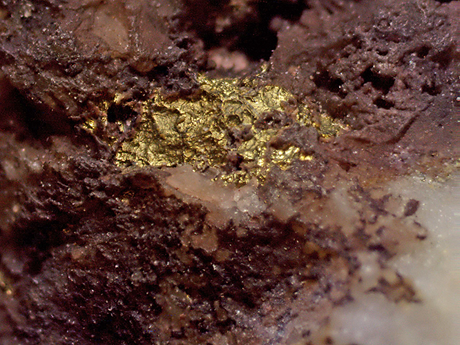 All that glitters… [By Pam Hamer]
Pam also brought several Zeiss accessories that are used with polarising microscopes and were formerly owned by Phil Robinson.
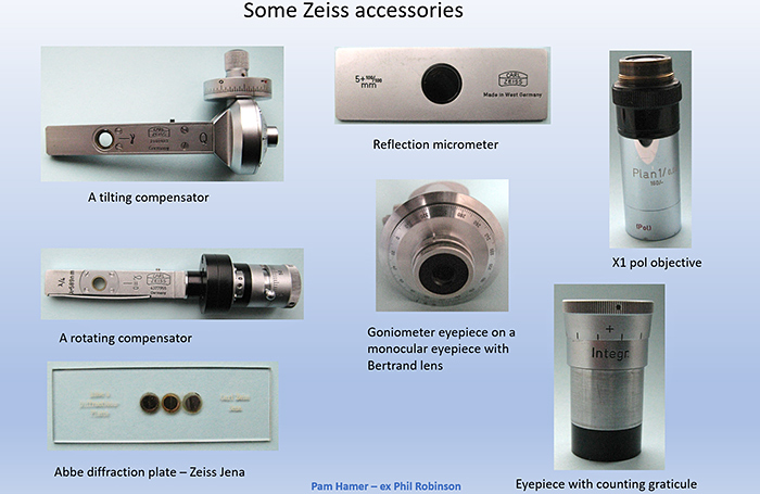 Zeiss polarising accessories [By Pam Hamer]
Terry Hope brought some samples from the pond in his garden, and used an inspection camera fitted with a zoom lens and mounted on a stand that provided transmitted light and a mechanical stage. The camera sent images via HDMI to a monitor. Robert Ratford lent Terry a trinocular Zeiss Axiostar Plus microscope with infinity-corrected Achrostigmat objectives, and he was able to use the same inspection camera. The specimens that we observed included filamentous algae, a gastrotrich, an ostracod, and lots of Paramecium.
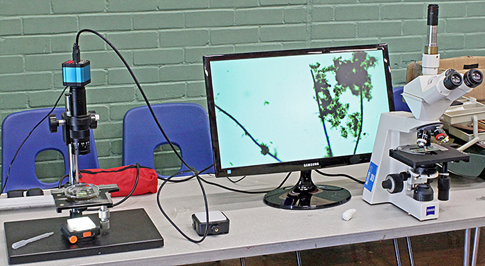 Terry Hope’s exhibit
 John Gregory, Robert Ratford (seated), Terry Hope and Mike Gibson
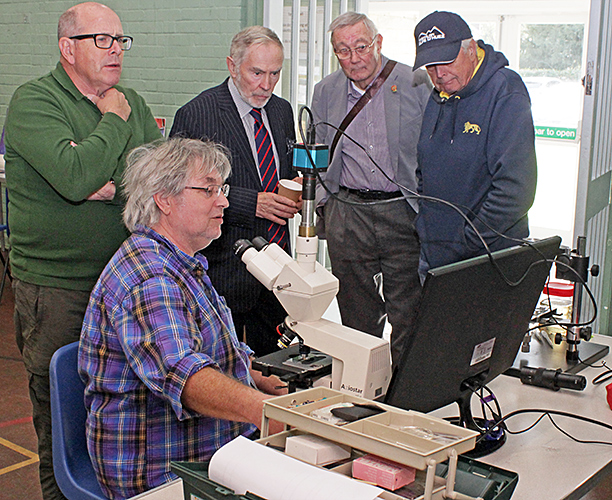
Michael Horwood brought slides that he has made and provided a Nikon stereomicroscope and a small AmScope compound microscope so that we could examine them. Some of them were ordinary specimens on standard size slides, all carefully ringed in various colours. Michael also brought lots of slides of wings of butterflies and moths that he has dried and mounted between pairs of slides (standard and double size) carefully sealed with copper tape. All of the wings came from old specimens where the bodies were decomposing.
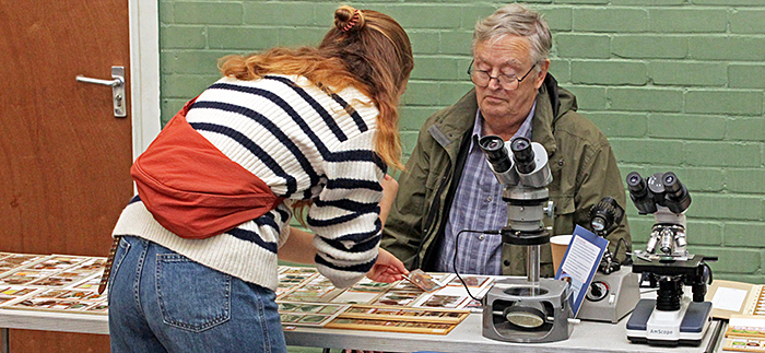 Michael Horwood with a visitor
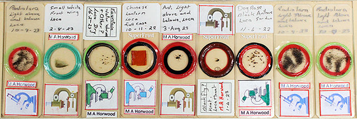 Michael Horwood’s slides
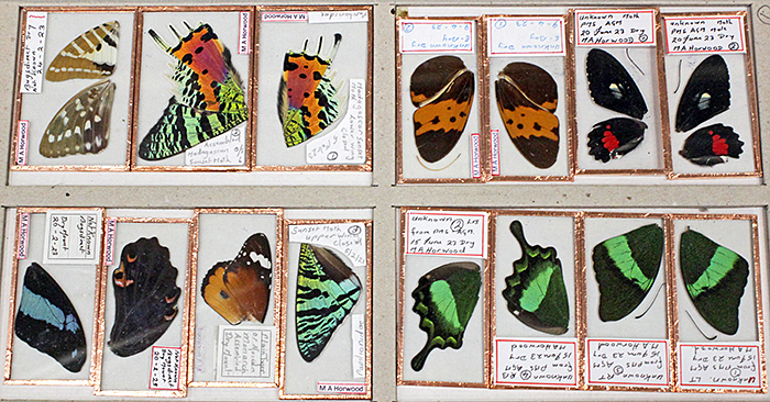 Michael Horwood’s slides of butterfly wings
Charles Hussey brought a Carl Zeiss inverted microscope, with some of its components resembling those familiar from upright Zeiss microscopes.
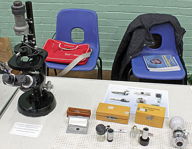 Zeiss inverted microscope and polarising accessories
Grenham Ireland brought a Zeiss Universal polarising microscope and a Zeiss IM35 inverted microscope equipped for phase contrast.
Specimens for the IM35 were mainly desmids from a pond in Dorset. They were held in a small plastic Cooper dish that Grenham had modified by cutting a circular hole in the base and cementing a coverslip over the hole. There was a Panasonic DMC-GF6 mounted via an adapter to the front camera port, sending images via WiFi to an old Tesco Hudl tablet and then via HDMI to a monitor.
Specimens for the Universal were slides of thin sections of metamorphic and igneous rocks. The camera was a 38MP FHD Camera V8 with a 0.5× reducing lens, sending images via HDMI to a monitor.
 Michael Horwood, Chris Thomas, Grenham Ireland and Stephen Edler
Graham Matthews brought several small monocular microscopes, mostly Lomo С11, that had been donated by The College of Richard Collyer in Horsham. Graham had refurbished as many of them as he could to make them available for the Club’s outreach activities.
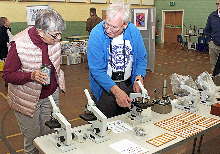 Pam Hamer and Graham Matthews
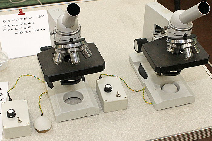 Refurbished Lomo microscopes for outreach
Graham also showed lots of slides that he has made of specimens from Warnham Local Nature Reserve.
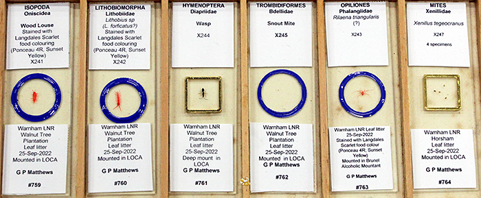 Graham Matthews’ slides
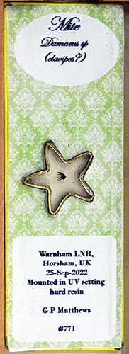 Star-shaped mount
Graham was the judge for the Eric Marson Award, so he also showed the slides that had been submitted, with photographs and photomicrographs.
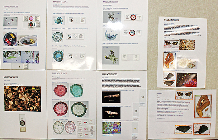 Slides entered for the Eric Marson Award
 PowerPoint presentation of the Eric Marson slides
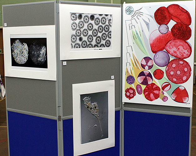
Robert Ratford showed the original artwork that he had submitted, all based on the drawing of a flea by Robert Hooke.
He also brought two trinocular stereomicroscopes and an inspection camera, and used them to show specimens including feathers, fabric, lichen and sycamore seeds.
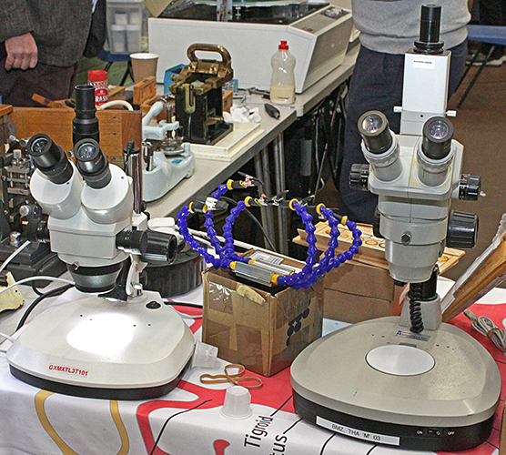 Stereo microscopes
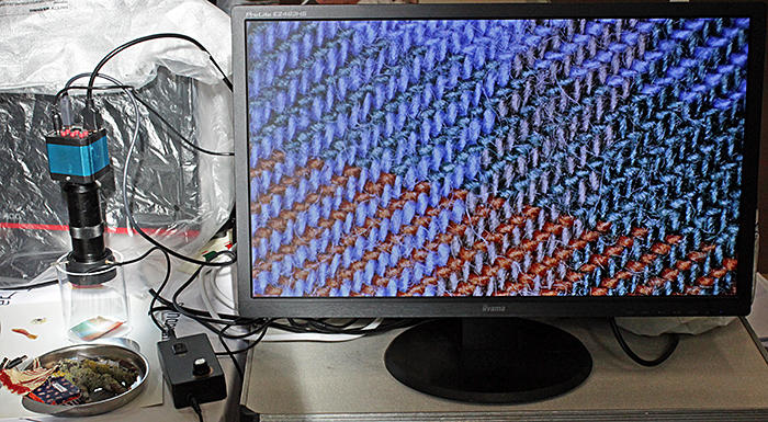 Inspection microscope
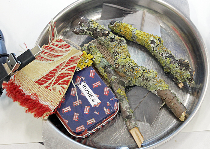 Some of Robert Ratford’s specimens
Robert also brought several journals and books, including Understanding Light Microscopy by Jeremy Sanderson, Contrast Techniques in Light Microscopy by S. Bradbury & P. J. Evennett, Understanding and Using the Stereomicroscope by Lewis Woolnough, Safe Microscopic Techniques for Amateurs: Slide Mounting by Walter Dioni, and The Microscope Made Easy by A. Laurence Wells.
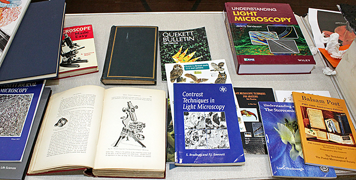 Books on microscopy
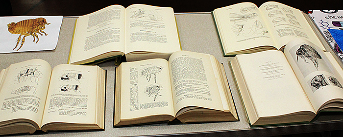 Books on fleas
Chris Thomas showed the original artwork that he submitted for the Exhibition and several of his paintings of pollen grains. Chris also brought some of the slides of pollen that he had made, and a trinocular Olympus BHA microscope with Plan objectives so that we could examine them. He used a USB-500 eyepiece camera with a 0.5× reducing lens sending images via USB to a laptop computer.
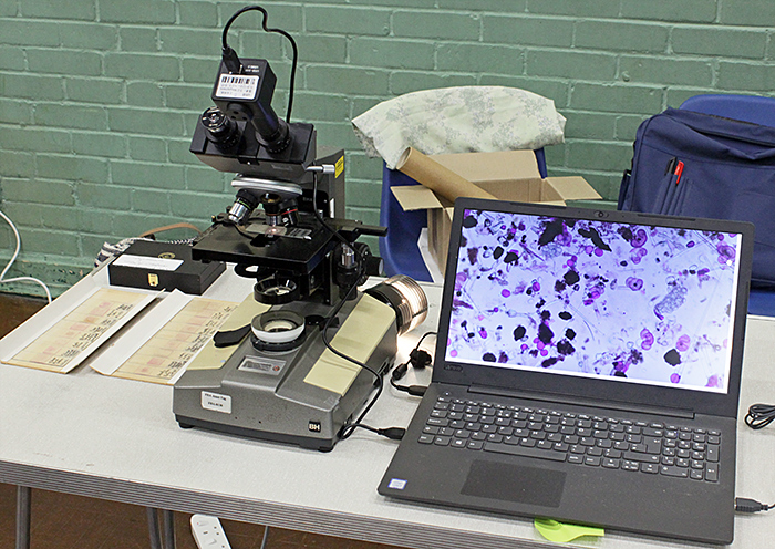 Chris Thomas’s exhibit
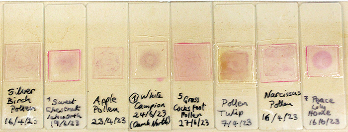 Chris Thomas’s pollen slides
Displays
Each year, we celebrate the work of a Quekett member. This year, David Linstead was chosen, and you can view a PowerPoint presentation about him, with lots of his photomicrographs. The PowerPoint and prints of some of David’s photomicrographs were displayed at the Exhibition.
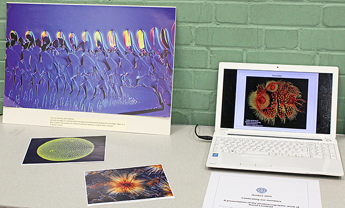 Celebrating our members – David Linstead
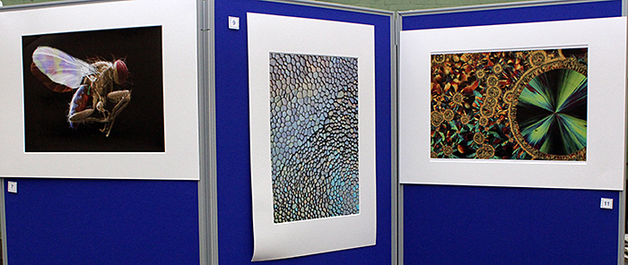
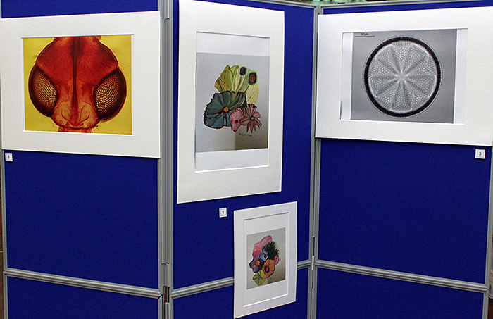
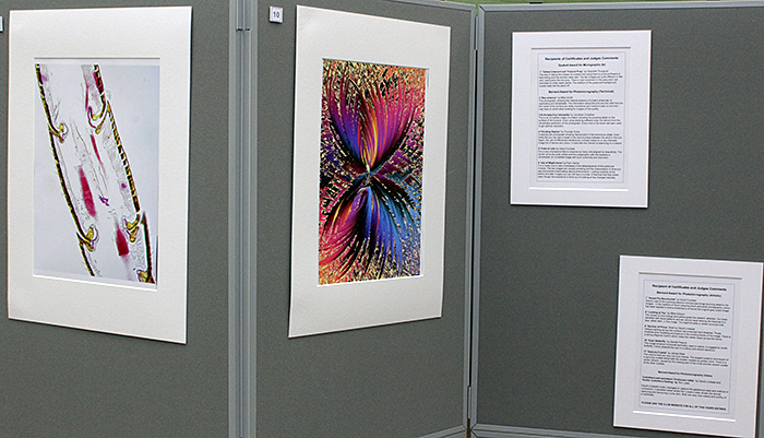
Shortly before the close of the meeting, Club President Terry Hope announced the names of the people who had been awarded certificates. The photomicrographs that received Barnard Awards and the slides that received Marson Awards were displayed at the Exhibition, together with photographs of the artworks by Gwyneth Thurgood that received a certificate. You can see all of the photographs and artworks that were submitted on these pages:
The slides that were entered for Eric Marson Awards were also displayed at the Exhibition:
Slides entered for the Eric Marson Award
PowerPoint presentation of the Eric Marson slides
You can see all of the slides that were submitted on this page:
Lectures
Quekex also features specialised lectures. This year two lectures were presented in person and via Zoom during the Exhibition. They were recorded, and Quekett members can watch them in the password-protected Members’ area.
“Fluorescence microscopy and its use in the life sciences” by Nicola Lawrence (Cambridge University)
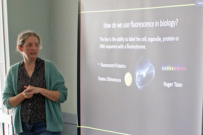 Nicola Lawrence
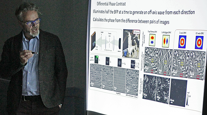 Kevin Webb
Acknowledgements
Our thanks to everyone who:
- brought exhibits and demonstrations
- submitted photomicrographs, videos, slides and artworks
- judged the photomicrographs, videos, slides and artworks
- booked the venue
- got out and packed away the tables and chairs
- organised the displays of photographs
- organised tea, coffee and biscuits
- arranged, broadcast and recorded the lectures
- publicised the event and the meeting report on social media
We hope to see you all again next year!
Report and most photographs by Alan Wood