Young Scientists’ Day
Saturday 27th June 2015
As in previous years, the Natural History Museum kindly allowed us to use the Angela Marmont Centre for our annual Young Scientists’ Day (part of the Club’s outreach programme) and provided a gazebo, tables and chairs near the pond in the Wildlife Garden. Among the visiting families, we were pleased to see familiar faces from the Wimbledon Common BioBlitz and Microscopes: More Than Meets the Eye.
James Rider used a net to collect material from the pond in the garden, and found plenty for us to observe under the gazebo and indoors. Specimens included lots of large waterfleas (Daphnia magna Straus) and phantom midge larvae (Chaoborus sp.), copepods, water boatmen (Notonecta sp.), water-lice (Asellus aquaticus (L.)), freshwater shrimps (Gammarus pulex (L.)), flatworms (including Dugesia lugubris (O. Schmidt)) and pond snails.
In the Angela Marmont Centre, Pam Hamer used two of the small battery-powered Natural History Museum Pocket Microscopes to show sugar crystals, salt crystals, paper, fabric, printing and beach microfossils mounted on plastic microscope slides, and she also had a stereomicroscope (for looking at pondlife and microwriting on banknotes, and checking £1 coins to see if they were forged) and copies of some of the Club’s leaflets.
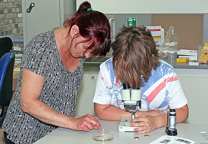 Visitors with Pam Hamer’s display
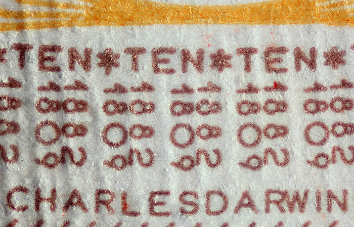 Microwriting on the reverse of a £10 banknote; the “D” in DARWIN is 0.4 mm tall
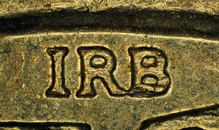 IRB – initials of designer Ian Rank-Broadley just below the Queen’s neck on a £1 coin; the letters are 0.65 mm tall
Norman Chapman brought some of his drawings and slides of pollen, and demonstrated how he extracts and prepares pollen.
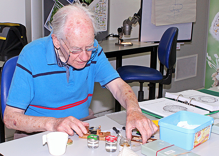 Norman Chapman
The highlight of the indoor display was the big television connected to the Club’s Canon EOS 550D camera and Zeiss Standard trinocular microscope, expertly demonstrated by David Linstead. This combination produced some stunning images of large waterfleas (Daphnia magna Straus), so that visitors could see their beating hearts, guts full of green algae, minute details of the cuticle and (with crossed polarising filters) the muscles. Crossed polars also allowed the muscles in the transparent larvae of phantom midges (Chaoborus sp.) to be clearly seen.
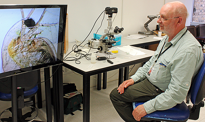 David Linstead, displaying Daphnia magna Straus on the Club’s television
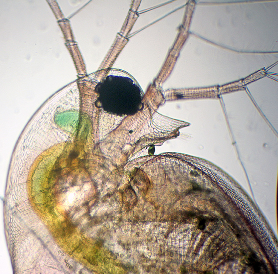 Head of Daphnia sp. [by David Linstead]
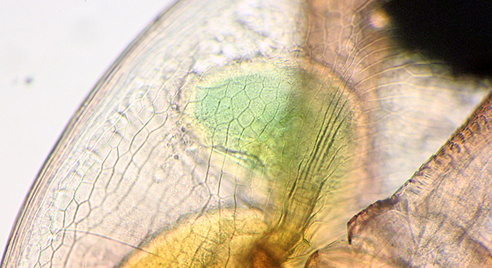 Blind caecum (green structure) in Daphnia sp.; the eye is at top right [by David Linstead]
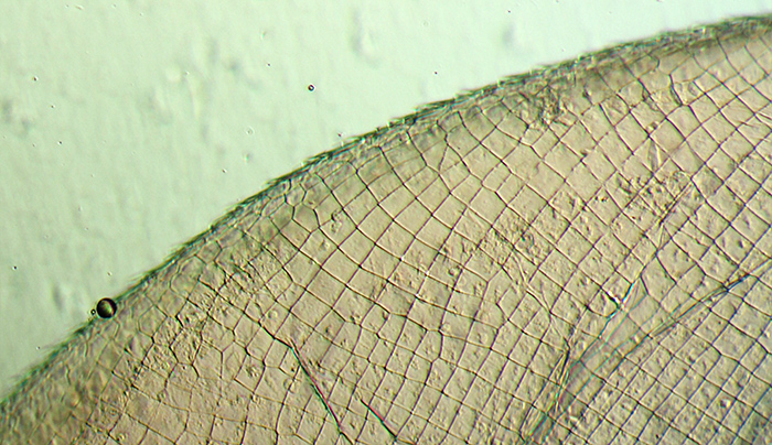 Cuticle of Daphnia sp. [by David Linstead]
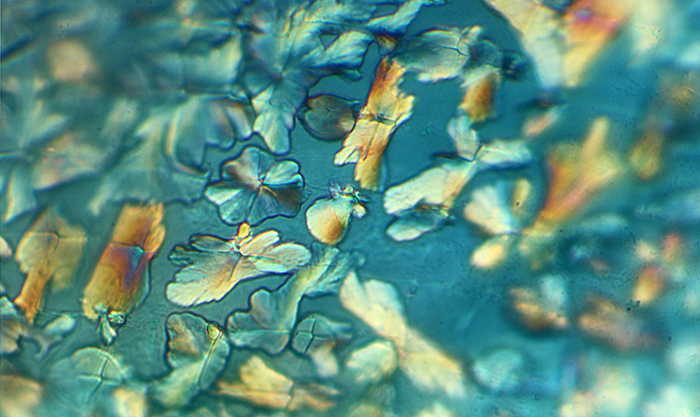 Crystals of calcite on cuticle of Daphnia sp. (polarised light) [by David Linstead]
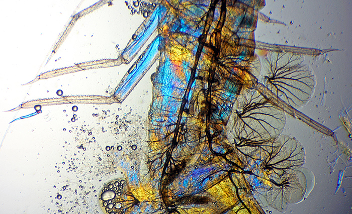 Gills and muscles of a mayfly nymph (using polarised light to make colours appear in the muscles) [by David Linstead]
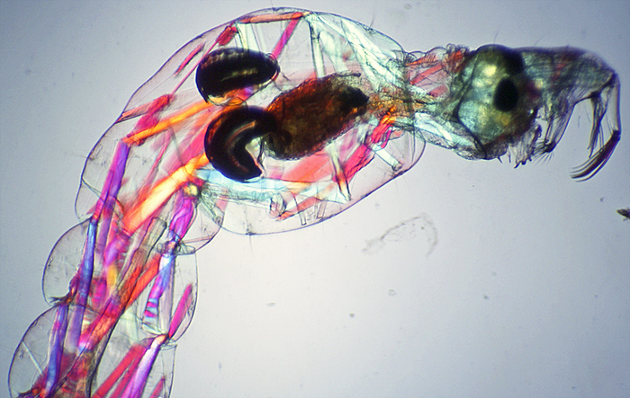 Front part of a phantom midge larva (Chaoborus sp.) (using polarised light to make colours appear in the muscles) [by David Linstead]
David also set up another of the Club’s Zeiss microscope with phase contrast, and the Club’s new trinocular stereomicroscope connected to the smaller television via David’s Sony NEX-5N digital camera and Scopetronix MaxView Plus for viewing pondlife.
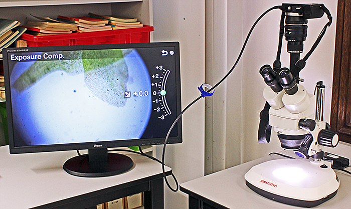 Stereomicroscope connected via a camera to a television
Alan Wood brought 2 of the new version of the Natural History Museum Pocket Microscopes (one with the attachment for viewing slides) and his small 20× stereomicroscope (from eBay seller gt-vision), a Helping Hand for holding specimens under the stereomicroscope, and a variety of specimens.
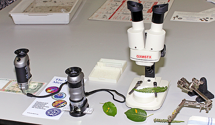 Natural History Museum Pocket Microscopes, a small stereomicroscope, and a Helping Hand supporting a lichen-covered twig
From his garden, he had fern fronds with circular sori underneath, pear leaves with a fungal gall, and lichen on a pear twig.
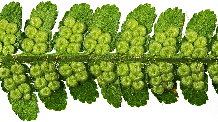 Circular sori (clusters of spore-containing sporangia) on the underside of a fern frond
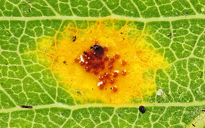 Early stage of gall caused by a fungus (Gymnosporangium sp.) on the top surface of a pear leaf (5 mm across)
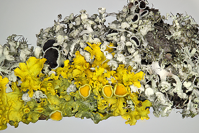 Yellow (Xanthoria sp.) and grey foliose lichens on a pear twig
Alan showed 2 types of colour printing, halftone used on the Quekett brochure, and offset litho used on a bank note from Peru, shown below at exactly the same magnification.
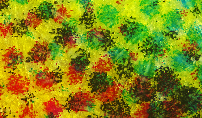 Halftone printing with dots of ink, used for the Quekett brochure
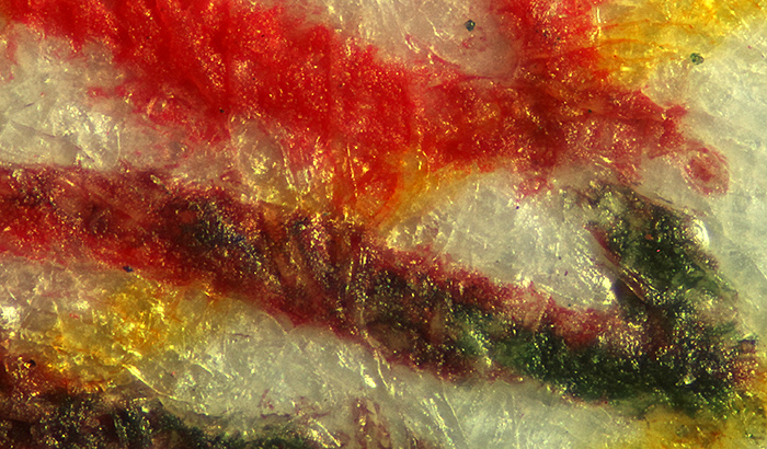 Offset lithographic printing, used for Peruvian banknotes
Alan also brought some slides to view with the NHM microscope, including a mosquito larva and a stained cross-section of the stem of a sunflower.
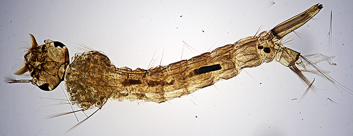 Larva of a mosquito (Culex sp.), on a prepared slide
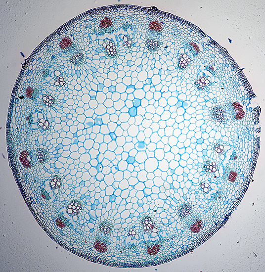 Stained cross-section of stem of sunflower (Helianthus annuus L.), on a prepared slide
Alan also showed unusual views of commonplace objects, a handkerchief (showing the interwoven white and blue threads) and the hooks on a piece of black Velcro®
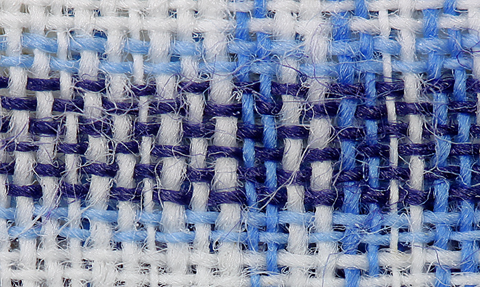 Blue and white handkerchief
 Black hooks of Velcro®
Outside in the Wildlife Garden, Paul Smith, Dennis Fullwood and Jenny Drewe showed pondlife using hand lenses, a Wild M11 compound microscope and a small 20× stereomicroscope (the same as Alan’s but from a different supplier).
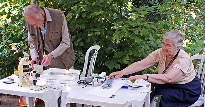 Dennis Fullwood and Jenny Drewe preparing the display under the gazebo
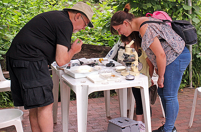 Paul Smith showing pondlife to visitors
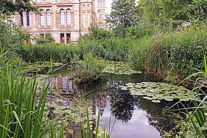 Pond in the Wildlife Garden of the Natural History Museum
Maurice Moss was there too, talking to visitors about pondlife and and showing them colours in Cellophane® using his polariscope. Carel Sartory, Nigel Williams and Jacky McPherson were also there.
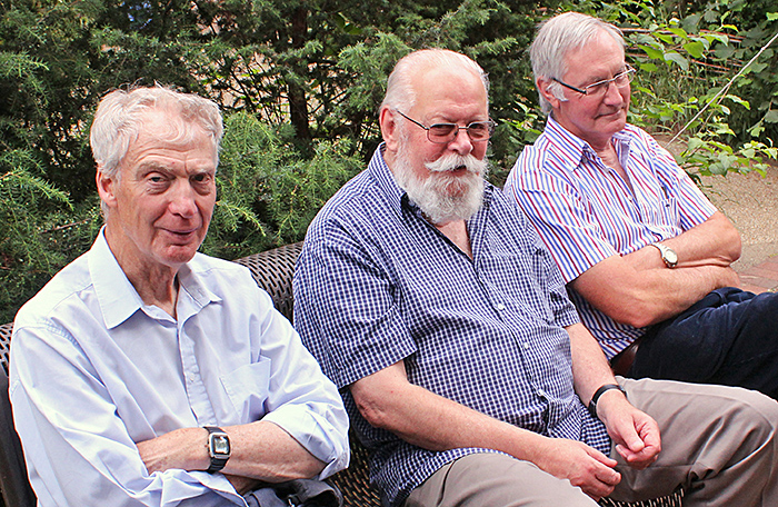 Left to right: James Rider, Carel Sartory and Nigel Williams
Report and photographs by Alan Wood, photomicrographs of live pond life by David Linstead

