

Photo of the Month
We have some very talented photographers in the Quekett, and there are hundreds of their images in the Photography showcase and the Slide showcase in the password-protected Members’ area. This page allows everyone to see some examples of their work, including photos that have received Barnard Awards at our Annual Exhibition of Microscopy. Copyright in these images belongs to the photographers, so please do not copy them without permission; if you contact the Club we can put you in touch with the photographer.
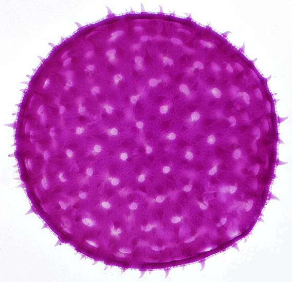
You can click on the latest Photo of the Month to see a full-size version.
May 2024
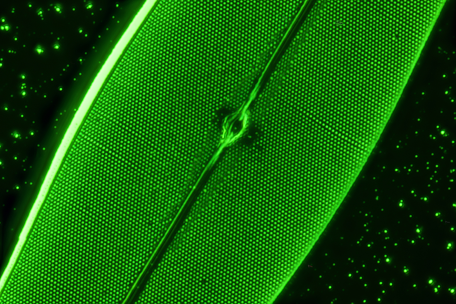 Pleurosigma angulatum diatom by John Dale.
Photographer: James Rider:
Subject: Aluminium-coated Pleurosigma angulatum diatom by John Dale.
Microscopy: Olympus BHS, 100× oil SPlanApo objective, 1.5× magnification changer, Aplanat Achromat Condenser and 550 nm green filter.
Photography: Canon EOS 6D Mark II (full-frame), ISO 100, operated by home-built stacking system. Stack of 65 photographs using Affinity Photo 2.
Reference: Balsam Post 40 12–14 (July 1998).
April 2024
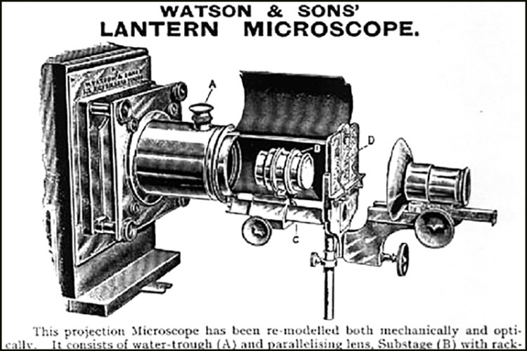 Lantern Microscope
An extract from a page of Watson’s Catalogue, shown at Quekex 2021
March 2024
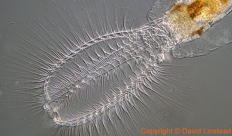
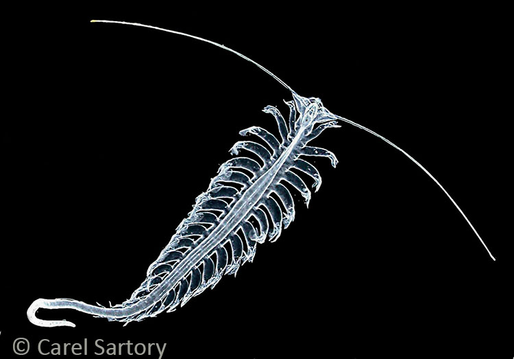 Tomopteris helgolandica
Photographer: Carel Sartory
Subject: Tomopteris helgolandica
Photography: A dark ground image of this planktonic polychaete often found in trawls taken at the marine microscopy weekend in Pembrokeshire. An unmounted but preserved specimen.
February 2024
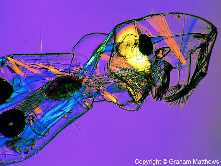
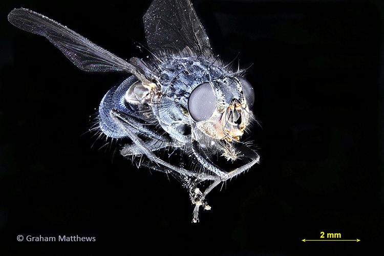 Housefly
Photographer: Graham Matthews
Subject: Adult housefly
January 2024
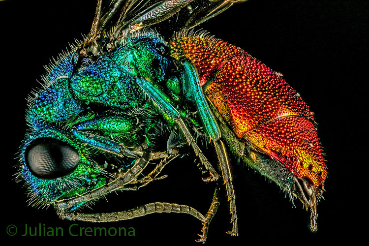 Ruby-tailed wasp
Photographer: Julian Cremona
Subject: Small parasitic wasp
Photography: Canon EOS 7D Mark II, 65 mm MP-E lens.
December 2023
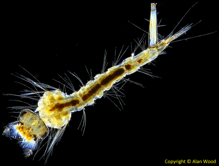
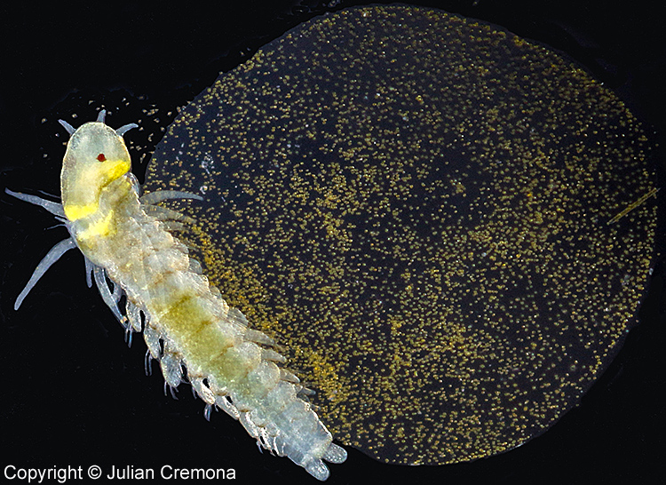
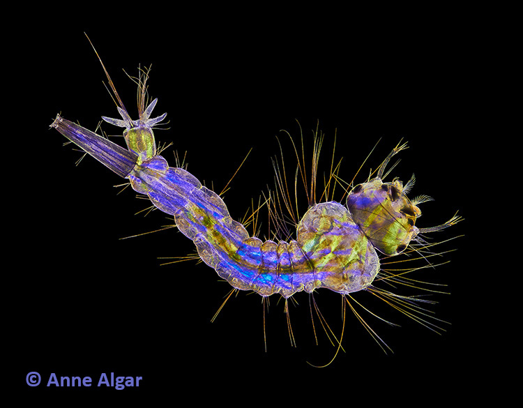 Mosquito larva
Photographer: Anne Algar
Subject: Mosquito larva
Photography: Olympus BHB microscope with 4× objective, polarizing and wave plate filters. Dark-field stacked image. Canon EOS 7D camera.
Anne received a Barnard Certificate of Excellence at the 2021 Quekex Annual Exhibition of Microscopy
November 2023
 Salicine crystal slide
Photographer: James Rider
Subject: Slide by J. G. Bradbury, paper-covered and appears to be vintage
Photography: Olympus BHS with 4× SPlanApo objective, Achromat Aplanat condenser and crossed polarizers. Full-frame Canon EOS 6D Mark II camera, ISO 100.
James received a Barnard Certificate of Excellence at the 2023 Quekex Annual Exhibition of Microscopy
October 2023
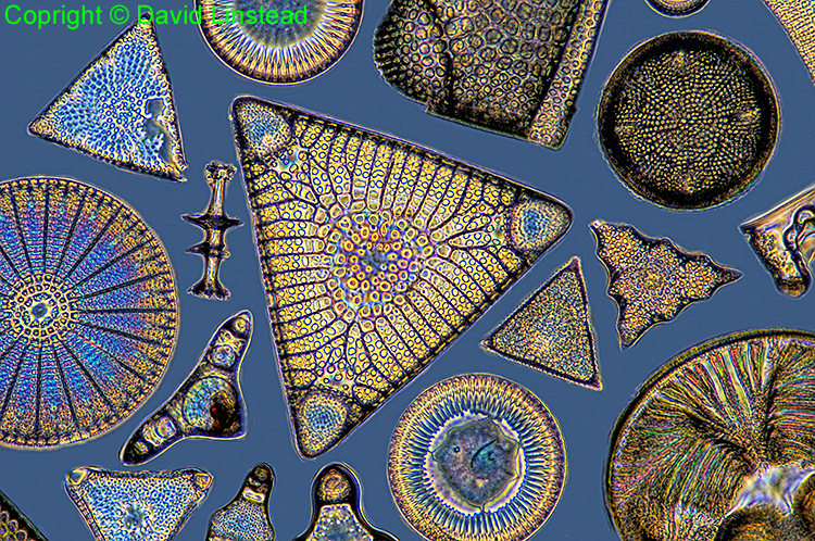
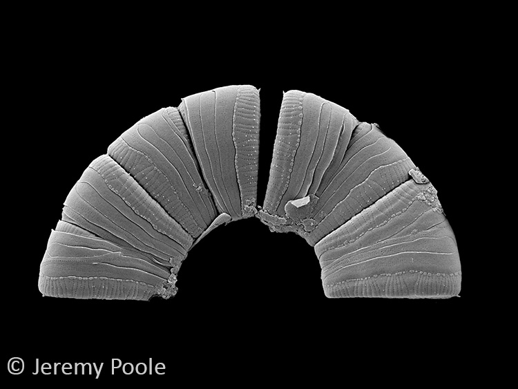 Meridion circulare diatoms
Photographer: Jeremy Poole
Subject: Meridion circulare diatoms in a semicircular formation.
Technique: Meridion circulare diatoms arrange naturally in a semicircular formation. This sample was collected near Malham Tarn, North Yorkshire and prepared, mounted on an SEM stub and imaged on a scanning electron microsope. The field width is 46.5 micrometers.
Photography: SEM. Post-processing to mask out unnecessary background and enhance brightness and contrast.
September 2023
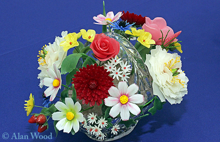 Beeswax flowers
Photographer: Alan Wood
Subject: Beeswax flowers at the 2022 Honey Show
July 2023
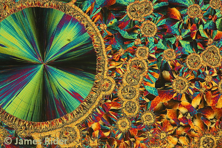
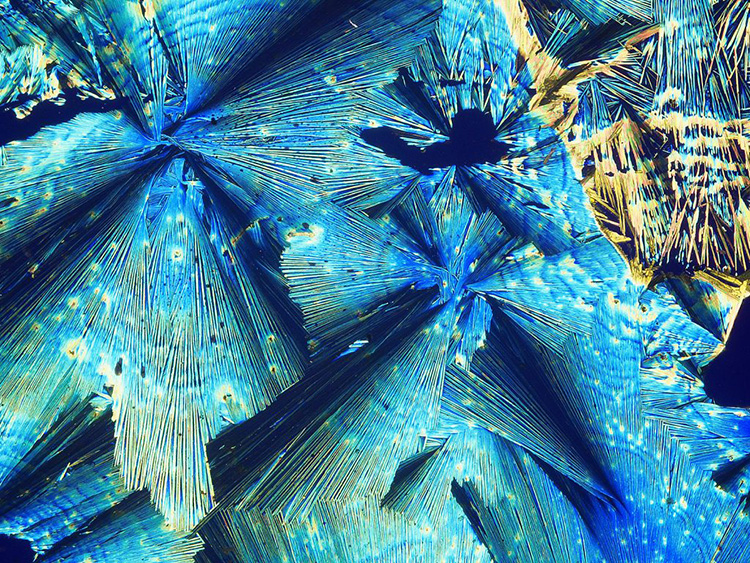
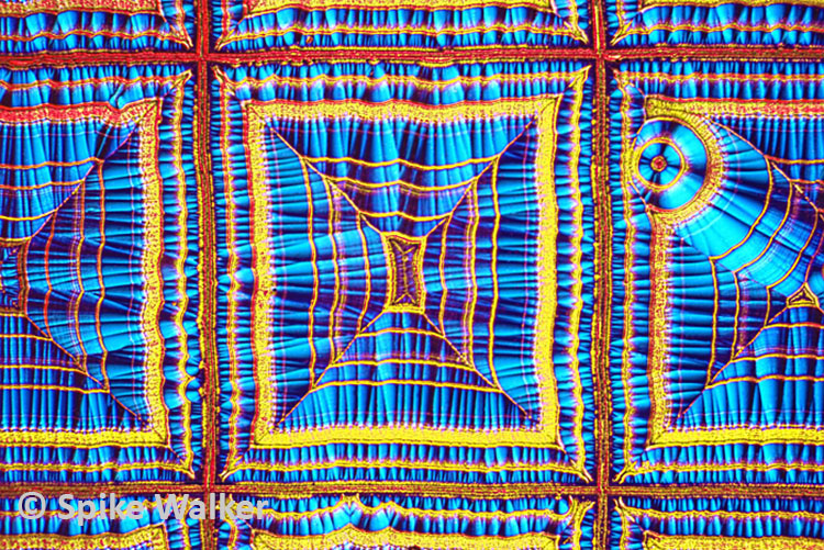 Vitamin C crystals, scratch preparation. Spikeberg
Photographer: Spike Walker
Subject: Vitamin C crystals
June 2023
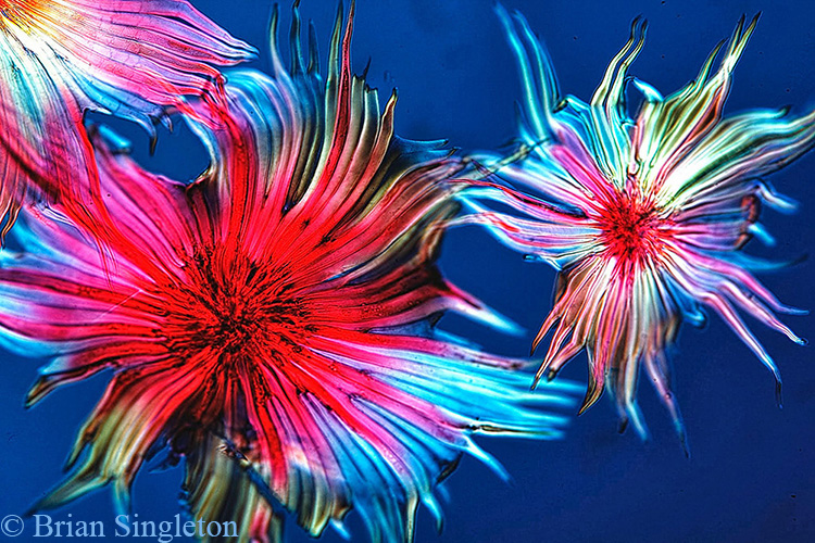 Leaf scales of Elaeagnus glabra (Thunb.)
Photographer: Brian Singleton
Subject: Leaf scales of Elaeagnus glabra(Thunb.) from a Biosil mount by John Wells
Technique: DIC with a Nikon E600 microscope, colours varied by altering the polariser setting.
May 2023
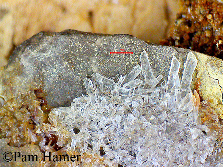 Rock Crystals
Photographer: Pam Hamer
Subject: Clear rock crystals
April 2023
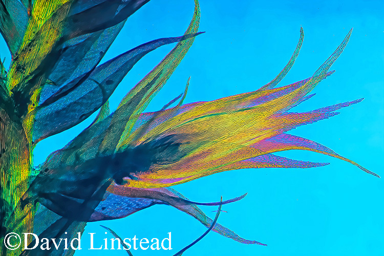 Side shoot of moss (?Mnium sp)
Photographer: David Linstead
Subject: Side shoot of moss, probably Mnium sp
Technique: subject flattened under coverslip and mounted in glycerol. Nikon Diaphot inverted microscope with 10× objective, polarised light and a retarder
Photography: Canon EOS 5D Mark II camera mounted on photoport of microscope. Stack of 40 images in Helicon Focus, Method B. Edited with Photoshop CS6.
February 2023
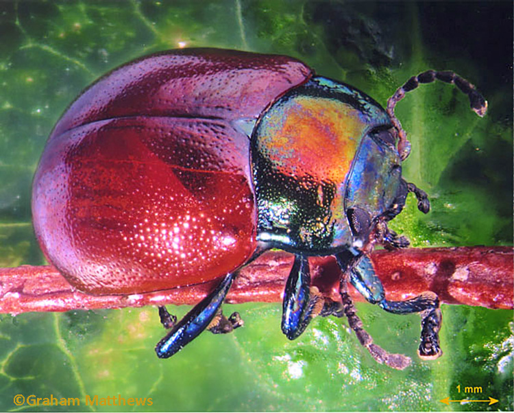 Beetle on a stick
Photographer: Graham Matthews
Subject: Chrysolina polita leaf beetle
Technique: A stack of 33 images combined in Helicon Focus Lite
Graham received a Special Merit Award at Quekex 2007 for this image
January 2023
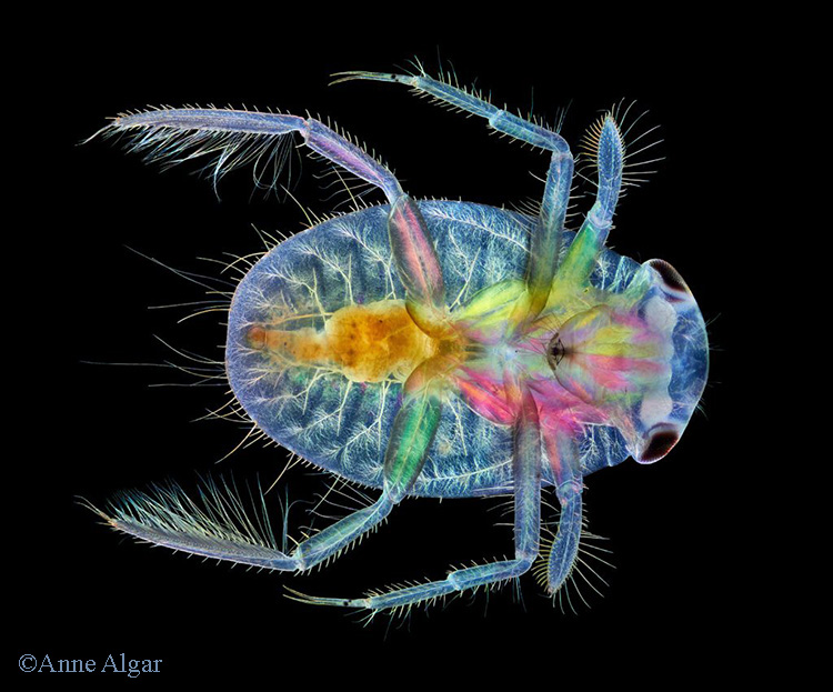 Water boatman
Photographer: Anne Algar
Subject: Water boatman showing internal and external structures
Technique: Olympus BHB microscope with 4X lens. Polarizing and wave plate filters, dark field.
Photography: Canon EOS 7D camera, image stacking
Anne received a Barnard Certificate of Excellence at the 2022 Quekex Annual Exhibition of Microscopy
December 2022
 Hazel catkin with pollen grains
Photographer: Mike Gibson
Subject: Part of a hazel catkin showing pollen grains
Technique: From a Biosil prepared slide. Gillet & Sibert conference microscope, x10 objective
Photography: Sony Cybershot W70 compact in afocal mode. Negative image from a stack of 15
November 2022
 Calcium platinocyanide crystals
Photographer: Carel Sartory
Subject: Calcium platinocyanide crystals – a 19th century preparation, maker unknown.
Technique: Wild M20 microscope, with 6× Wild Pan Fluotar objective and Olympus NFK 2.5× photo eyepiece. Köhler illumination, crossed polarisers.
Photography: Olympus OMD E-M10 mark II mirrorless digital camera.
Carel received a Barnard Award Certificate of Excellence at the 2021 Quekex Annual Exhibition of Microscopy.
September 2022
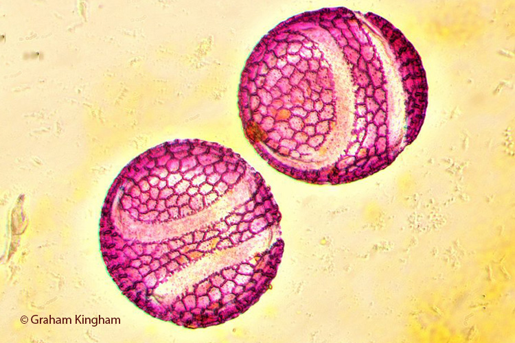 Passionflower pollen
Photographer: Graham Kingham
Subject: Passionflower pollen grains
Technique: Stained with fuchsin, Meiji ML 2000 microscope, brightfield illumination
Photography: ToupTek USB3 E3 CMOS camera, stacked images
Graham received a Barnard Award Certificate of Excellence at the 2021 Quekex Annual Exhibition of Microscopy.
August 2022
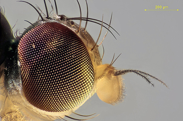 Head of a small fly
Photographer: Graham Matthews
July 2022
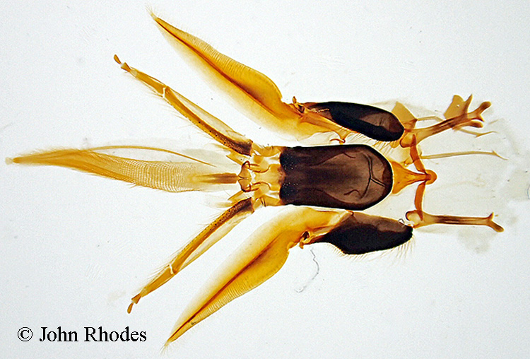 Mouthparts of Apis mellifera
Photographer: John Rhodes
Subject: Mouthparts of the European Honeybee (Apis mellifera)
Technique: Olympus TG-4 camera on microscope setting
June 2022
 Stinging hair on stem of nettle
Photographer: Alan Wood
Subject: Stinging hair on stem of nettle (Urtica dioica L.)
Technique: Olympus SPlan 4× objective, NFK 2.5× photo eyepiece, Canon EOS 5D Mark II camera, stack of 24 images in Zerene Stacker, levels adjusted in Adobe Photoshop Elements.
Alan received a Barnard Award Certificate of Excellence at the 2018 Quekex Annual Exhibition of Microscopy
May 2022
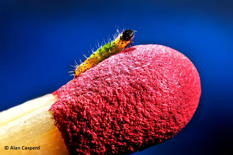 Hatchling of cabbage white (Pieris brassicae) on head of a match.
Photographer: Alan Casperd
April 2022
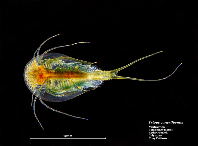 Tadpole shrimp (Triops cancriformis)
Photographer: Tony Pattinson
Subject: Temporary mount of a tadpole shrimp.
Technique: Ventral view, specimen mounted in cedarwood oil.
Tony received a Barnard Award Certificate of Excellence at the 2020 Quekex Annual Exhibition of Microscopy.
March 2022
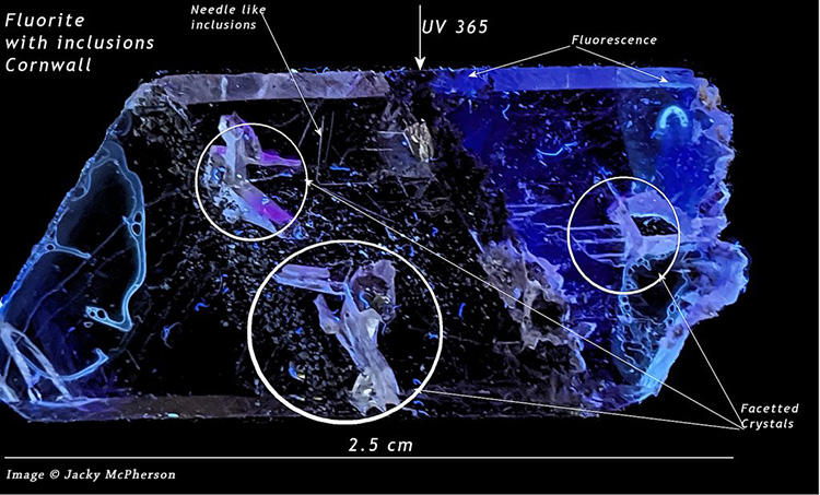 Fluorite crystal, UV illumination
Photographer: Jacky McPherson
Subject: Slide of uncovered slice of fluorite crystal with inclusions (25×10mm, 1.5mm thick) from Cornwall, by J. A. Bottomley.
Technique: Oblique transmitted illumination from UV torch with a 365nm band-pass filter to reduce glare.
Photography: Google Pixel 3a smartphone hand-held and braced on pile of boxes. Corel Paintshop Pro XVIII to crop the image, raise the black, reduce artefacts and add annotations.
Jacky received a Barnard Award Certificate of Excellence at the 2020 Quekex Annual Exhibition of Microscopy
February 2022
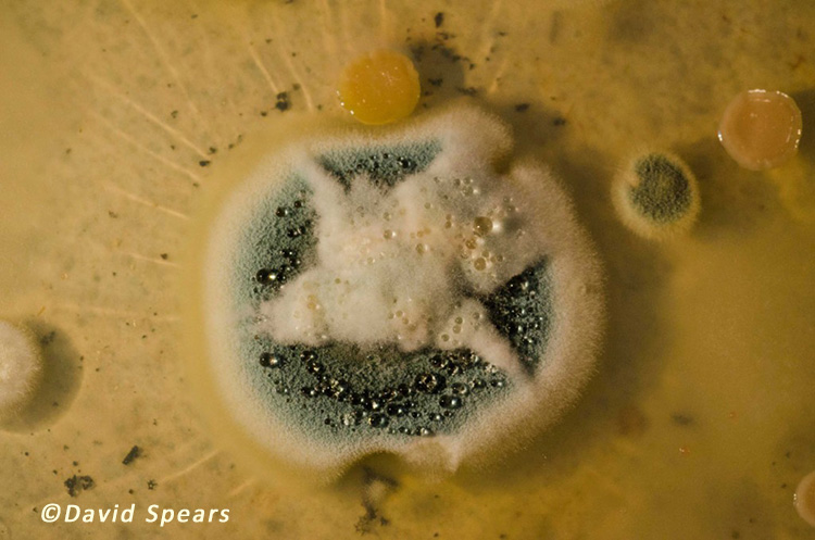 Mature Penicillium colony
Photographer: David Spears.
Subject: Mature Penicillium colony.
Technique: This is the end shot of a time-lapse sequence shot for a TV programme. The nutrient agar was seeded with a tiny amount of the original strain of Penicillium used by Alexander Fleming in his development of antibiotics. The culture dish was warmed and contained in a humid chamber during growth. Camera was Nikon D7000 with a 55mm Micro Nikkor lens.
January 2022
 Bladderwort
Photographer: Julian Cremona
Subject: Bladderwort (Utricularia sp.) showing the underwater bladder for catching prey.
Technique: Composite stack of 38 images, 4× magnification.
December 2021
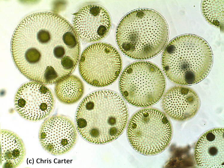 A group of Volvox aureus colonies
Photographer: Chris Carter
Subject: Group of Volvox aureus colonies
Equipment: Olympus CX41 with semi-apochromatic objective, bright field. Paxcam 3 camera.
Chris received a Barnard Award Certificate of Excellence at the 2020 Quekex Annual Exhibition of Microscopy
November 2021
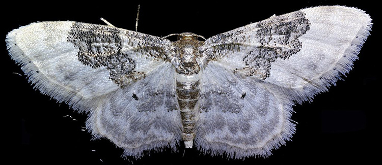 Least carpet moth (Idaea rusticata) – live specimen
Photographer: Chris Thomas
Subject: Live least carpet moth (Idaea rusticata). The insect stayed absolutely still for over an hour during the photography process.
Equipment and Processing: Reichert Zetopan stand. The image is the result of over 1500 photos taken with a 10× objective projected directly onto the camera sensor without an eyepiece, combined into 90 focus stacks and stitched using Hugin.
October 2021
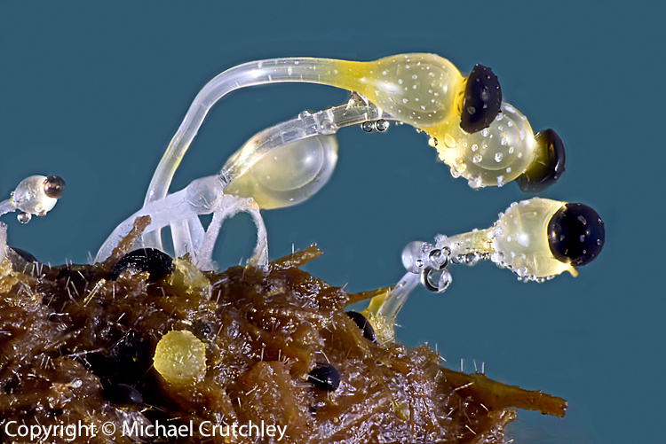
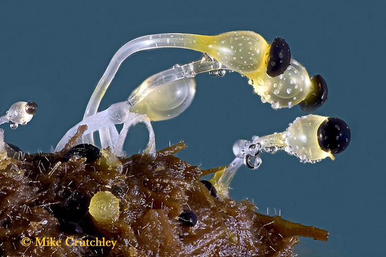 Pilobolus sp. on dung, showing fruiting bodies
Photographer: Mike Crutchley
Subject: The fungus Pilobolus sp. on dung, showing fruiting bodies. Nicknamed ‘the dung cannon’ because it can shoot the spore capsule some distance to aid dispersal.
Mike received a Barnard Award Certificate of Excellence at the 2016 Quekex Annual Exhibition of Microscopy
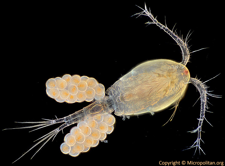
September 2021
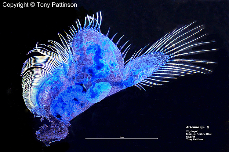
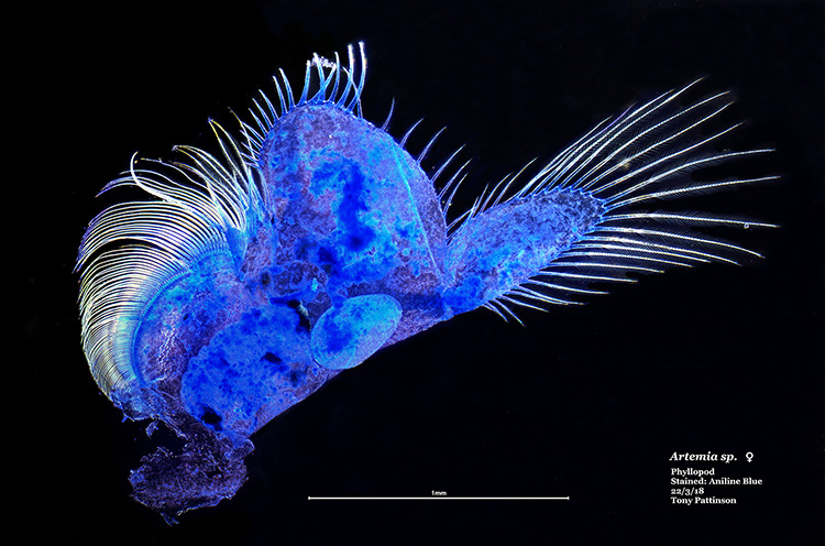 Female brine shrimp phyllopod
Photographer: Tony Pattinson
Subject: Phyllopod of female brine shrimp (Artemia sp.), stained with aniline blue. Scale bar is 1 mm.
August 2021
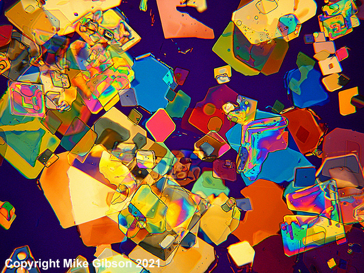 Borax crystals (2021 RMS Microscience)
Photographer: Mike Gibson
Subject: Borax crystals
Equipment: Wild M20 microscope, x19 objective
Polarised light
Mike’s image won joint 1st prize in the Light Microscopy Physical Sciences category at the 2021 Microscience Microscopy Congress
July 2021
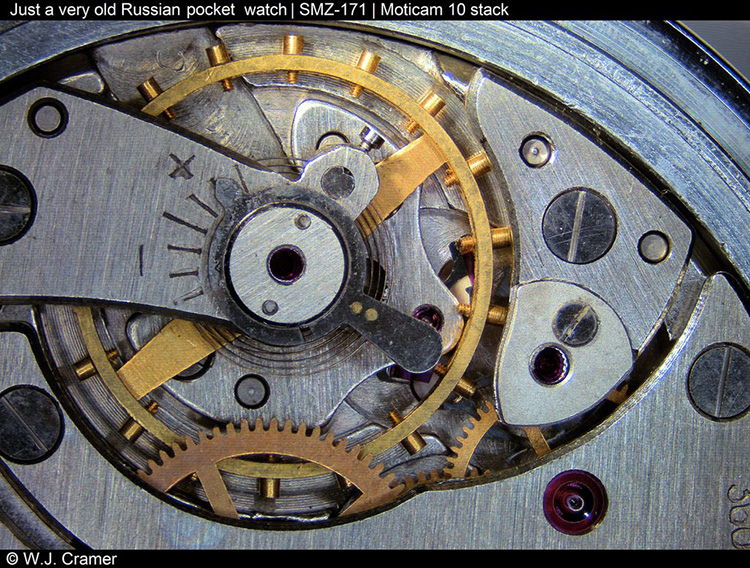 Old Russian pocket watch
Photographer: Willem Cramer
Subject: A very old Russian pocket watch
Equipment: Motic SMZ171 stereomicroscope
Camera: Moticam 10
Stacked image
March 2021
 Coronal funnel of Stephanoceros fimbriatus
Photographer: David Linstead
Subject: Coronal funnel of the sessile rotifer Stephanoceros fimbriatus
Equipment: Zeiss Standard microscope, 16× Reichert fluorite objective, DIC illumination. Canon EOS M3 camera afocally coupled to the microscope using Leitz Periplan 10×20 threaded eyepiece.
Software: Stack of 44 images in Zerene Stacker, PMax, edited with Photoshop CS6.
November 2020
 Fossil diatoms from Oamaru
Photographer: David Linstead
Subject: Arrangement of fossil Oamaru diatoms by Steve Beats. The diatomite deposit at Oamaru in New Zealand dates from the late Eocene to the early Oligocene period.
Equipment: Leitz Ortholux microscope, Zeiss ×16 Neofluar phase objective, Heine condenser, phase contrast plus polarisers (phacopol)
October 2020
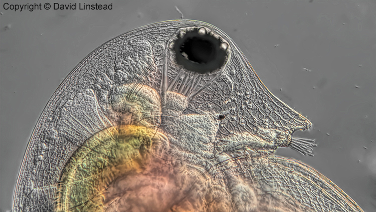 Daphnia head and shoulders portrait
Photographer: David Linstead
Subject: Upper body of Daphnia pulex, live specimen anaesthetized with lidocaine.
Equipment: Zeiss Standard microscope, 16× Reichert fluorite objective, DIC illumination. Afocal coupled Panasonic GX80 camera.
Software: Stack of 23 images in Zerene Stacker, PMax. Edited with Photoshop CS6.
January 2019
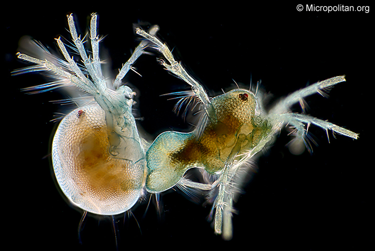 Mating mites
Photographer: Wim van Egmond
Subject: Mating water mites (Arrenurus sp., family Hydrachnidae). Water mites are freshwater arachnids. Note the sexual dimorphism. Magnification ×44 when printed 10 centimetres wide.
Technique: dark-ground illumination
December 2018
 Phyllopod of Artemia
Photographer: Tony Pattinson
Subject: Phyllopod (swimming appendage) of female brine shrimp (Artemia sp.). Stained with aniline blue. Scale bar is 1 mm.
Technique: dark-ground illumination
February 2018
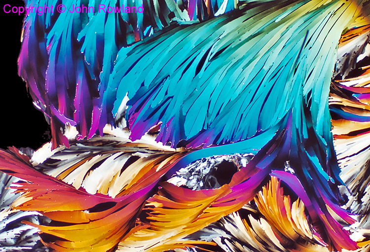 Hexadecane crystals
Photographer: John Rowland
Subject: Low-melting-point wax (hexadecane) crystallised on slide
Technique: crossed polarisers, 4× objective
January 2018
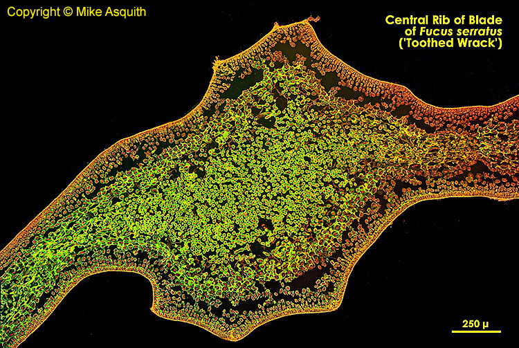 Toothed wrack
Photographer: Mike Asquith
Subject: Central rib of blade of Fucus serratus (toothed wrack)
Technique: Fluorescence
December 2017
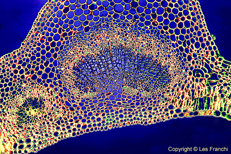 T. S. leaf vein of lilac
Photographer: Les Franchi
Subject: T. S. leaf vein of lilac (Syringa sp.), oblique Rheinberg illumination, field of view ~ 0.9 mm.
November 2017
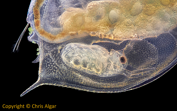 Baby Daphnia inside its mother
Photographer: Chris Algar
Subject: Daphnia from garden pond.
Equipment: Canon EOS 7D Mark I camera, Nikon 10× finite objective (without photo eyepiece), stack of 62 frames, 1/6 sec, ISO 200.
Software: RAW images processed with Capture One and exported as 16 bit TIFFs, then stacked using Zerene Stacker, minor retouching in Photoshop CS5.
May 2017
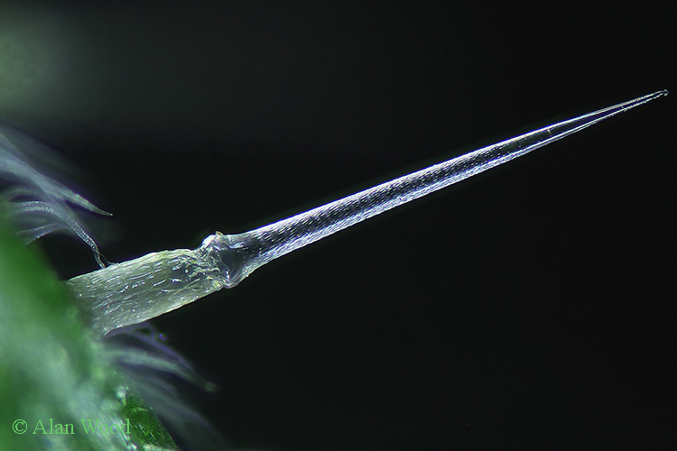
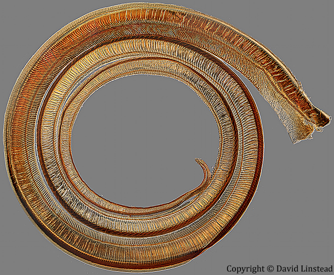
 Larva of Culex pipiens L.
Photographer: Alan Wood
Subject: Larva of Culex pipiens L. prepared without pressure, slide by T. Gerrard & Co.
Equipment: Canon EOS 40D camera with 60 mm EF-S macro lens, single exposure at f/11, home-made dark-ground illuminator with inverted 144 LED ring-light.
Software: Adobe Photoshop Elements 11 to adjust levels, clean the black background and sharpen.
March 2017
 Cyclops sp.
Photographer: Wim van Egmond
Subject: Copepod, Cyclops sp., dark ground stacked image
February 2017
 Moth tongue
Photographer: David Linstead
Subject: Moth tongue (proboscis), slide by Charles Morgan Topping
Equipment: Zeiss Standard 18 microscope, ‘mismatch’ DIC, Zeiss Jena ×6.3 0.20 apochromat objective, ×16 objective prism inserted upside down, and the DIC condenser prism in the ‘I’ position. Canon EOS M camera, afocal coupling.
Software: Stitch of 43 images with Microsoft ICE
January 2017
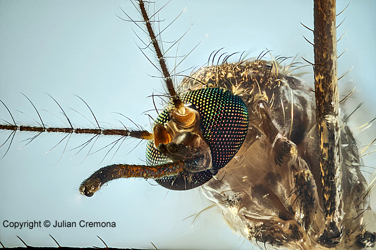 Head of mosquito
Photographer: Julian Cremona
Subject: Head of a mosquito
Equipment: Vickers 10× objective mounted in a body cap attached to a series of extension tubes and a Canon EOS 7D Mark II. Magnification on sensor approximately 20×. Lighting was a twin macro flash. A StackShot was used to take a set of 190 images that were then stacked.
December 2016
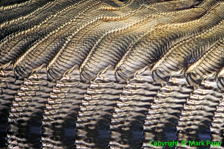 Radula of periwinkle
Photographer: Mark Papp
Subject: Palate (radula) of a periwinkle (marine gastropod mollusc, family Littorinidae) from an old slide.
Equipment: Wild M20 compound microscope with crossed polarisers, Plan Fluotar objective and Olympus 3.3× photo eyepiece, Canon EOS 700D camera.
Software: Stack of about 15 images.
November 2016
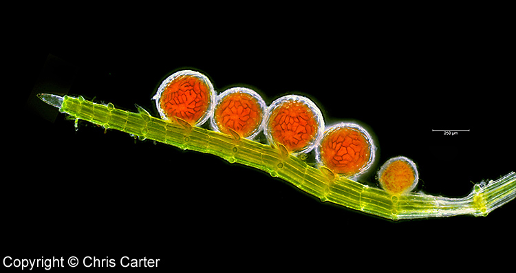 Antheridia of Chara fragifera
Photographer: Chris Carter
Subject: A row of antheridia (male reproductive organs) of Chara fragifera Durieu, a rare stonewort from Cornwall that has separate male and female plants.
Equipment: Olympus CX41 compound microscope with ×10 semi-apochromatic objective, dark-ground illumination. C-mount PAXcam3 USB camera with Paxit image capture software.
Software: The image is made up of 30 sub-images: 5 horizontal stitched sections, each at 6 focus levels stacked with Helicon Focus. Necessary processing with Adobe Photoshop CS6.
October 2016
 Fruiting bodies of Pilobolus on dung
Photographer: Mike Crutchley
Subject: The fungus Pilobolus crystallinus (F. H. Wigg.) Tode on cow dung, showing fruiting bodies. This fungus is nicknamed ‘the dung cannon’ because it can shoot the spore capsule some distance to aid dispersal of the spores.
September 2016
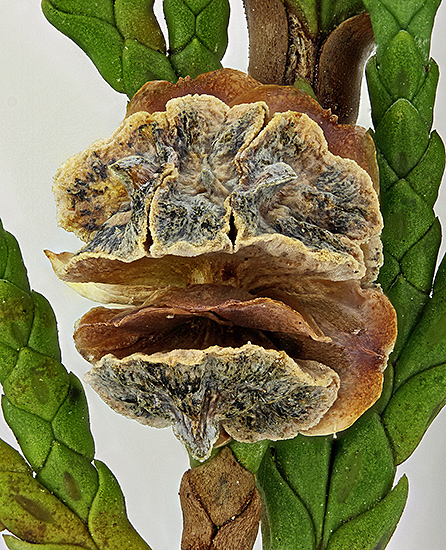 Seed cone of Lawson’s cypress
Photographer: Alan Wood
Subject: Seed cone of Lawson’s cypress (Chamaecyparis lawsoniana (A. Murray) Parl.); the cone is about 5 mm wide.
Equipment: Canon EOS 600D with 60 mm EF-S macro lens + Olympus 13 cm supplementary lens, lighting was 2 CF bulbs through a Kleenex diffuser.
Software: EOS Utility, Zerene Stacker (to combine 95 images), Photoshop Elements 11 (to adjust levels and sharpen)
August 2016
 Phantom midge larva (Chaoborus sp.)
Photographer: Graham Matthews
Subject: Musculature of phantom midge larva (Chaoborus sp.).
Technique: Crossed polarisers plus a retarder to add colours to the muscles, which are normally colourless.
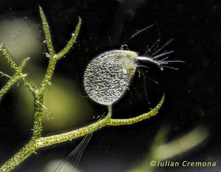
July 2016
 Larval paddleworm on Phaeocystis colony
Photographer: Julian Cremona
Subject: Larva of a paddleworm stuck to the surface of a colony of the diatom Phaeocystis globosa Scherffel. These diatoms form a colony by secreting a ball of gelatinous slime with large numbers of diatoms over the surface. Taken at the Dale Fort Marine Microscopy Weekend in May 2016.
Equipment: Canon EOS 7D Mark II attached with several extension tubes to a 65mm MPE lens held vertically. The MPE lens was set on 5× magnification and with the extension tubes this would make it around 7×. Canon Twin macro flashes were used one above and one below the specimen but offset. A black card was below the stage so a translucent dark background resulted. The digital image was cleaned up with some cloning of detritus and slight adjustment of levels and curves in Lightroom.
June 2016
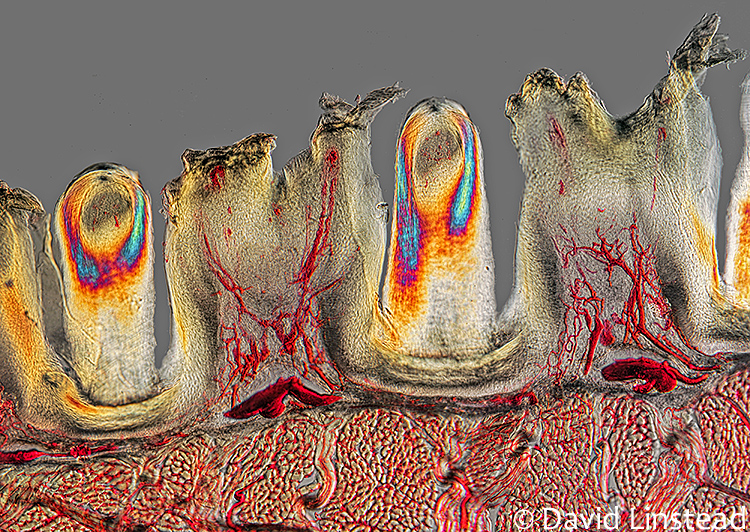 Papillae on the surface of a cat tongue
Photographer: David Linstead
Subject: Two different types of papillae on the surface of a cat tongue, from a Victorian slide sold by Watson. Injected blood capillaries, longitudinal and circular muscle blocks and keratinised papillae are all clearly shown.
Equipment: DIC using a Wild ×10 0.45 Fluorite objective with a Nikon ×10 DIC prism on a Nikon Diaphot. Stitch of 26 images. Canon EOS 40D camera.
May 2016
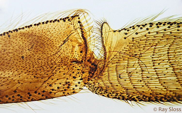 Corbicula of a bee
Photographer: Ray Sloss
Subject: The corbicula (or ‘pollen basket’ and ‘pollen press’) on the hind leg of a bee, from a slide made by Dennis Fullwood at Flatford Mill Field Centre in February 2016.
Equipment: Leitz PL ×6 objective and a ×2.5 relay lens, stack of 10 images.
April 2016
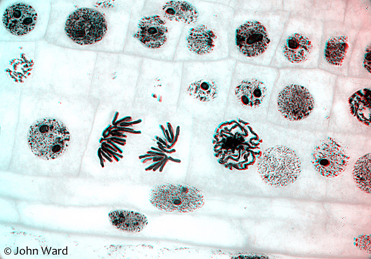 Chromosomes in root tip of onion
 ![]()
How to view: To see this anaglyph 3D image properly, you need red/cyan glasses with red for your left eye. Suitable glasses are available cheaply on eBay.
Photographer: John Ward
Equipment: Zeiss ×100/1.25 objective and DIC
March 2016
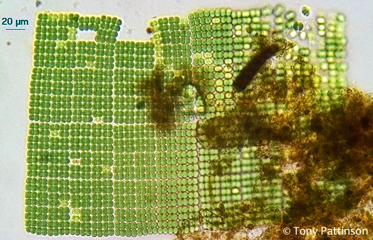 Merismopedia elegans A. Braun ex Kützing
Photographer: Tony Pattinson
Specimen: Merismopedia elegans A. Braun ex Kützing, rectangular colonial cyanobacteria found and photographed during the July 2015 Quekett excursion to the Basingstoke Canal.
Equipment: Olympus A 20× objective, 4× CTS eyepiece, 3 MP ScopeTek C-mount camera with a 25 mm Cosmicar lens mounted afocally.
February 2016
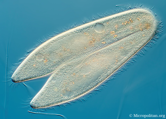 Conjugating Paramecium – sex amongst the Protozoa!
Photographer: Wim van Egmond
DIC image showing conjugation in the protozoan Paramecium, a sexual phenomenon in which paramecia of compatible mating types fuse temporarily and exchange genetic material. During conjugation, the micronuclei of each conjugant divide by meiosis and the haploid gametes pass from one cell to the other. The gametes of each organism then fuse to form diploid micronuclei. The old macronuclei are destroyed, and new ones are developed from the new micronuclei.
January 2016
 Mallow pollen grain
Photographer: Alan Wood
Subject: Stained single grain of pollen of mallow (Malva sp.) from an NBS slide, diameter 150 µm
Equipment: Olympus SPlan 40× objective, NFK 2.5× photo eyepiece, Canon EOS 5D Mark II
Software: EOS Utility, Zerene Stacker (to combine 13 images), Photoshop Elements 11 (to adjust levels and sharpen)