

Wimbledon Common Nature Club
Sunday 7th January 2024
The Wimbledon Common Nature Club is run by Auriel Glanville, helped by Luci Teuma and two teenage assistants (Alexander and Oliver Mallett). The Club welcomes children from 6–14 years old to come and discover the world of nature on Wimbledon Common. They meet for 2 hours each month in the Information Centre, the same venue as used by Quekett members on excursions, the Weekend of Nature and the Open Day.
As part of the Quekett’s outreach programme, Paul Smith, Barry Wendon and Alan Wood took an assortment of microscopes and some interesting slides and specimens to show the children at the “Life Under a Microscope” meeting. As usual, some of the parents were keen to have a look too.
When everyone had arrived, Auriel led us on a walk through the grassland and woodland so that the children could collect specimens to take back to the Centre. There had been a lot of rain recently, so there were lots of puddles on the paths and in the grass.
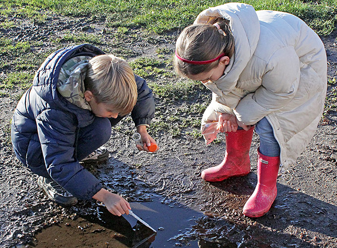 Collecting samples from a puddle
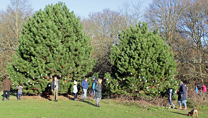 Christmas trees
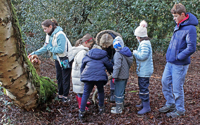 Collecting in the woods
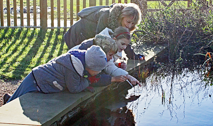 Collecting from the new pond (watched by Luci Teuma)
Back in the Centre, we started looking at the specimens that the children collected during the walk.
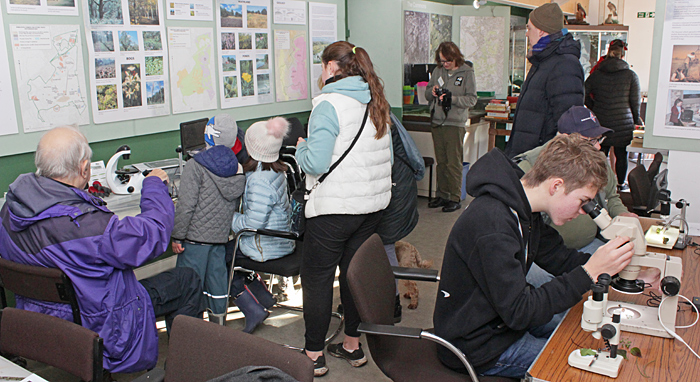 Children and adults in the Information Centre
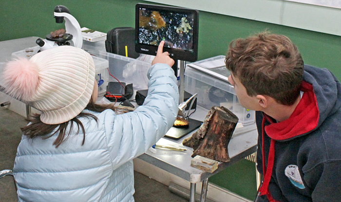
Barry Wendon brought his new digital microscope that has built-in lamps and a screen, and used it to show lichen and other specimens.
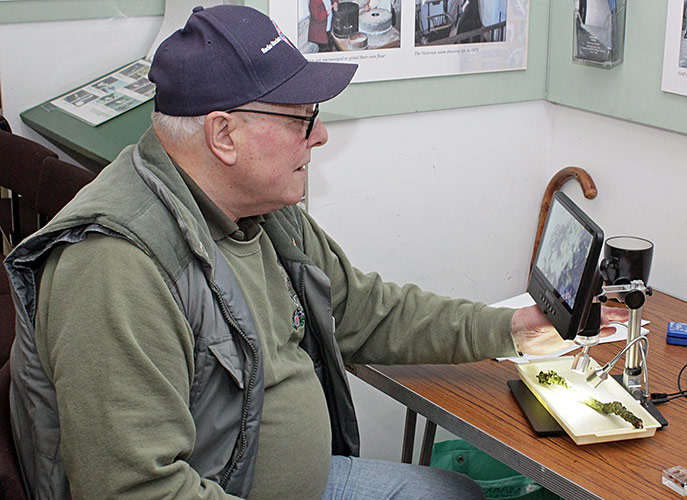 Barry Wendon with his digital microscope
Paul Smith brought his small Telmu inverted microscope that is good for looking at aquatic specimens from below. He collected some samples from the cattle trough and found some filamentous algae, possibly Cladophora sp. It was covered in diatoms, wedge shaped with a curve, attached at the thin end, possibly Rhoicosphenia sp.
 Inverted microscope
Paul also brought his Tomlov DM602 digital microscope. This includes a screen and lamps, and comes with three interchangeable lenses to provide a wide range of magnifications, and an accessory that provides transmitted light for viewing slides. Paul’s specimens included a beetle, a hornet and a scorpion embedded in resin blocks. He also brought some star sand from a beach in Okinawa (Japan), where the grains of sand are made of calcium carbonate and are the empty shells of foraminifera. The children enjoyed looking at the specimens that they collected on the walk, including bark, moss and bracket fungi.
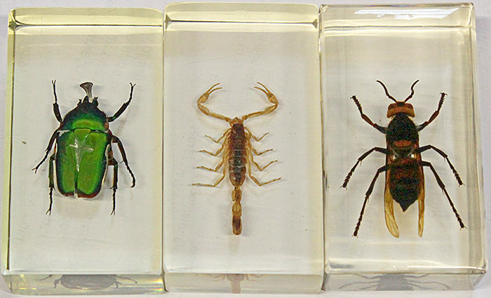 Insects and a scorpion in resin
 Tomlov DM602 digital microscope
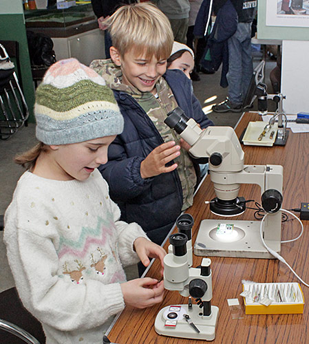
Alan Wood brought his Olympus SZ4045 stereo microscope, fitted with an LED ring-light for looking at opaque specimens and an LED stage plate for looking at transparent specimens such as microscope slides. Alan brought some lichen, a parakeet feather, a butterfly wing and several microscope slides. During the walk, he collected various leaves, galls and seed heads.
 Stereo microscopes
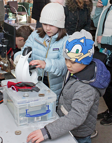
Alan also brought a small stereomicroscope (the STX Stereo Microscope sold on Amazon by GT Vision).
Here are some of the specimens as seen under a microscope:
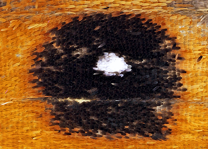 Eye-spot of a small heath butterfly (1.2 × 1 mm) (Coenonympha pamphilus)
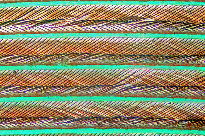 Feather of a ring-necked parakeet
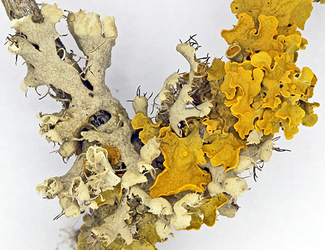 Grey and yellow lichens on a small twig
Most specimens on microscope slides need to be very thin, so that light can pass through them. Small ones (like a flea, a foraminiferan or a hydra) can be mounted whole. Larger specimens (like a grasshopper, a rabbit embryo, or the bud or stem of a plant) usually need to be sliced into thin sections. The sections are so thin that they are almost colourless, so they are usually stained to make them easier to see. Large specimens such as a caterpillar or a beetle can be made thin by removing their insides and then flattening them.
Here are some of the microscope slides:
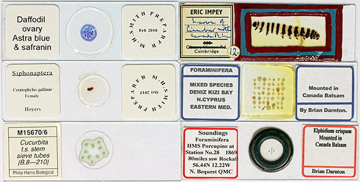 Microscope slides
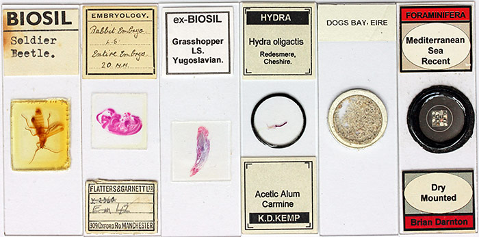 Microscope slides
And here are some of the specimens on the microscope slides, mostly photographed through a microscope:
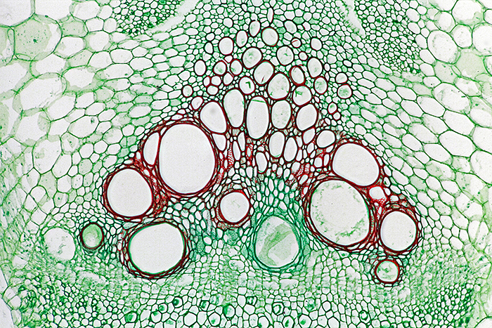
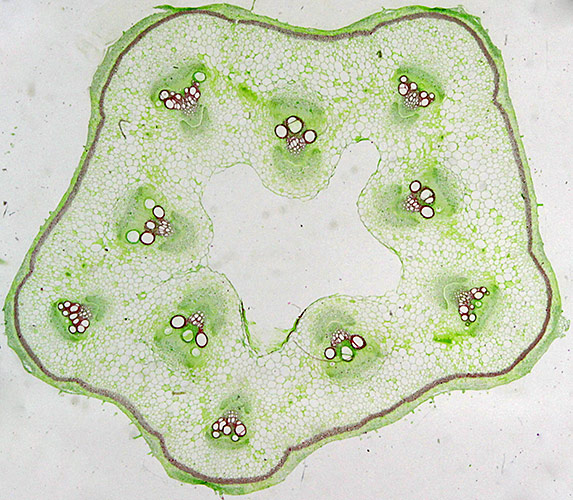 Stained section of stem of Cucurbita sp., showing 10 groups of sieve tubes
 Sieve tubes in stem of Cucurbita sp. (bottom right in photo above)
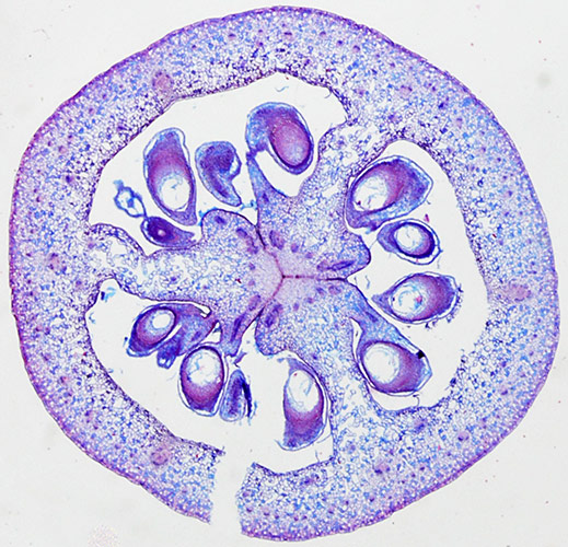 Stained section of ovary of daffodil
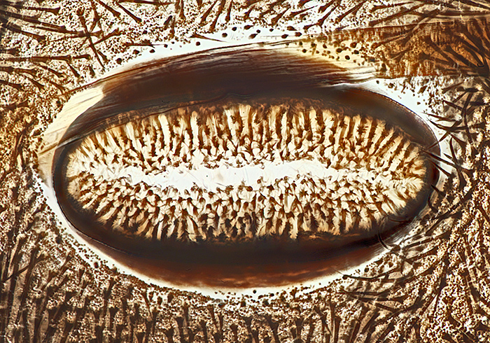
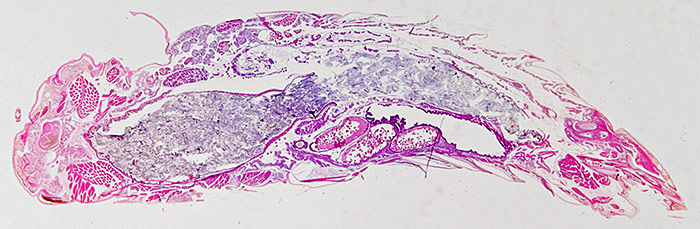 Stained longitudinal section of grasshopper
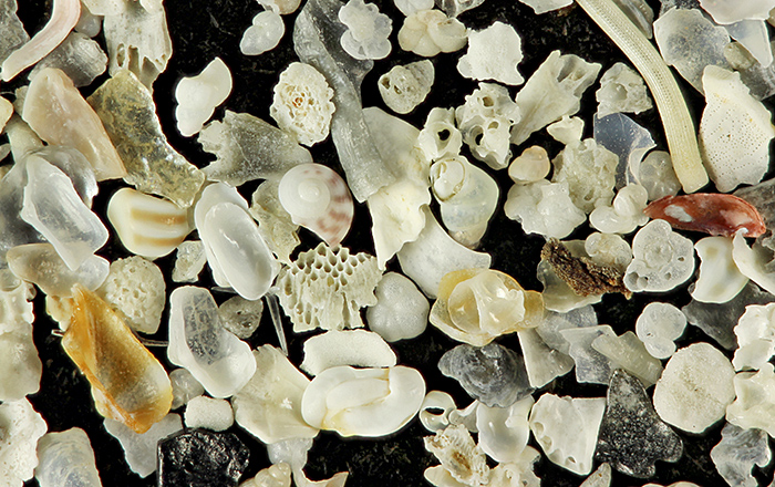 Shell sand from Dog’s Bay on the west coast of Ireland
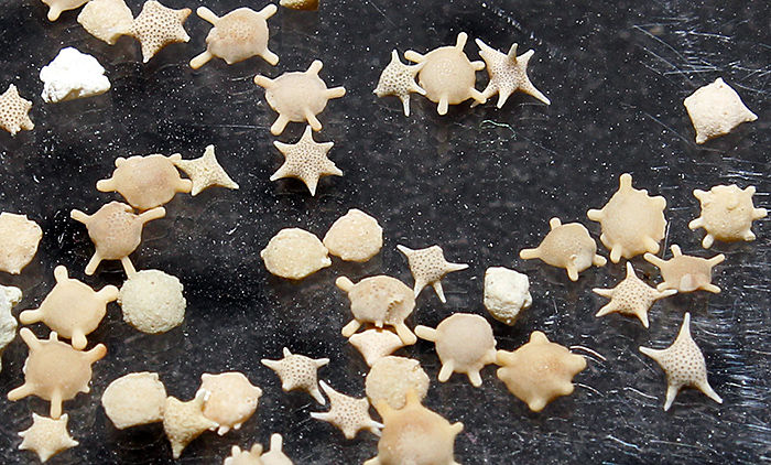 Star sand from Okinawa, Japan
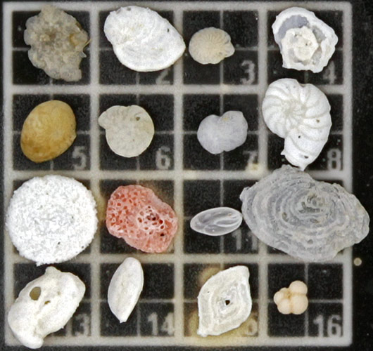 Sixteen forams arranged on a 5×5 mm grid
 Spiracle of caterpillar of cinnabar moth (0.2 mm) (Tyria jacobaeae) (the slide has the complete skin of the caterpillar)
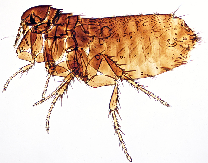 European chicken flea (Ceratophyllus gallinae) (2.6 mm)

Stained longitudinal section of a rabbit embryo (14 mm)
One of the parents kindly took a group photograph for us:
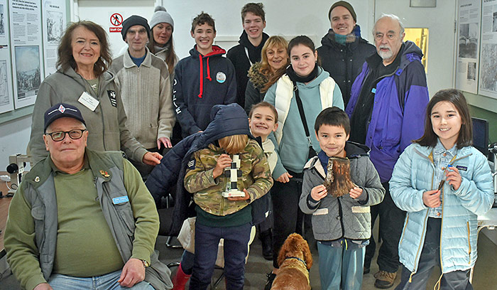 Group photograph
Report and most photographs by Alan Wood