

Workshop on contrast enhancement
Saturday 14th February 2026
Chris Thomas opened the workshop in the Angela Marmont Centre in the Natural History Museum with a presentation on microscopy that included several methods of enhancing contrast without needing to use stains: dark-ground and Rheinberg illumination, crossed polarisers, phase contrast and differential interference contrast. The last two need expensive equipment, but Chris set out to show that the others can be implemented at minimal cost.
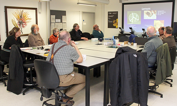 Chris Thomas’s presentation
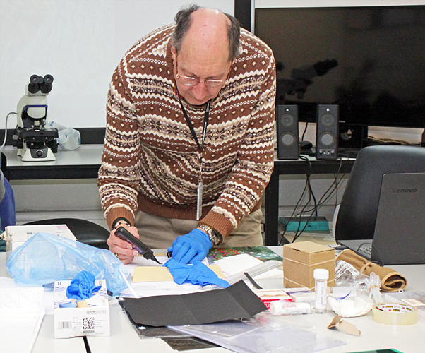 Chris Thomas demonstrating
For specimens, Chris provided potato starch, toilet tissue, dog hair, down feathers and flight feathers. Starch gives colourful images with crossed polarisers. Toilet tissue is designed to separate into cellulose fibres when soaked in water, and the fibres show colours with crossed polarisers and retarders.
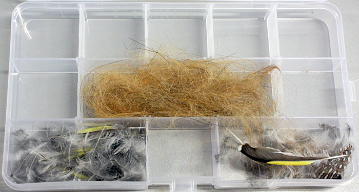 Fur and feathers
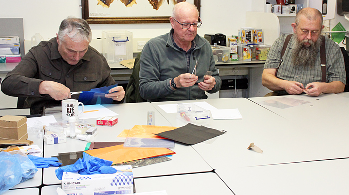
For making slides, Chris provided slides, coverslips and UV-setting resin. For making dark-ground stops, he provided sheets of thin black card. For making Rheinberg filters, he provided sheets of red, blue, green, orange and yellow plastic film. For crossed polars, he provided sheets of polarising material and clear plastic films. Chris also provided pipettes, scissors, rulers and clear adhesive tape.
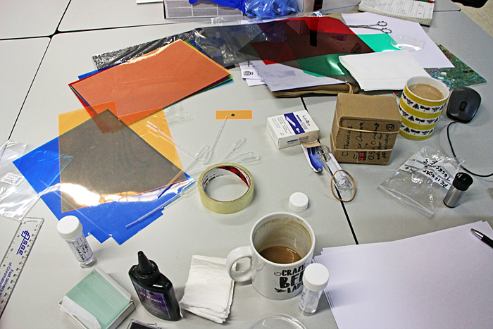 Coloured films and other materials
Dark-ground stops are usually circular, but Chris showed us that octagons also work and are easy to make; cut a square and then cut off the four corners. Rheinberg filters are also usually circular, but Chris showed us that opposing squares can also give interesting results.
 Dark-ground stop and Rheinberg filter
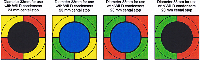 Complex Rheinberg filters made by Carel Sartory
We were able to use the Museum’s Olympus CX41 and CX43 microscopes which are equipped for phase contrast, and their condensers also have positions for dark-ground and normal bright field. Polarisers and retarders can be placed over the light outlet, where they are easy to rotate. The binocular heads are secured, so we placed analysers on top of our slides. Dark-ground stops and Rheinberg filters need to be as close as possible to the iris diaphragm in the condenser, so Chris showed us how to improvise using dark-ground stops on glass slides or Rheinberg filters on strips of plastic films that we held just below the condenser.
In addition to regular members, we were pleased to welcome Jonathan Palmer to his first workshop and four new members, Jim Archer, Laurence Peacock, Melissa Thompson and Remigiusz Lecbyl.
 Laurence Peacock, Jonathan Palmer and Remigiusz Lecybyl
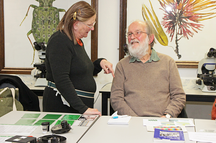 Jacky McPherson and Jim Archer
 Members gossiping
Here are photomicrographs of two of the sample specimens:
 Down feather using dark-ground illumination (10× objective)
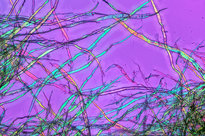 Teased tissue using crossed polarisers plus retarder (10× objective)
 Teased tissue using crossed polarisers plus retarder (10× objective)
Here are some photomicrographs from the Quekett archives, showing the sort of images that can be produced with contrast enhancement:
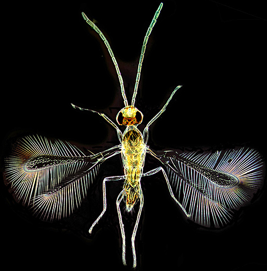 Fairy fly using dark-ground illumination [by Lewis Woolnough]
 Stonewort using dark-ground illumination [by Chris Carter]
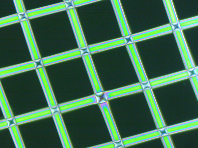
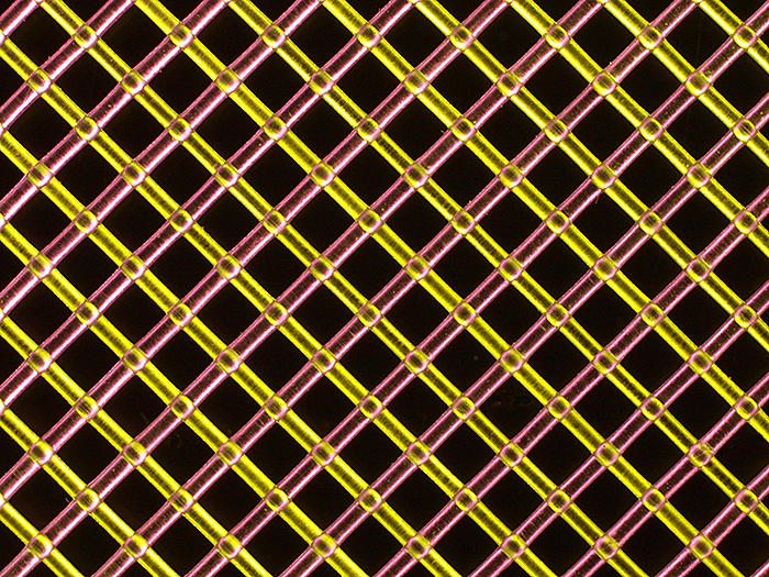 Colourless polyester mesh using Rheinberg illumination [by Carel Sartory]
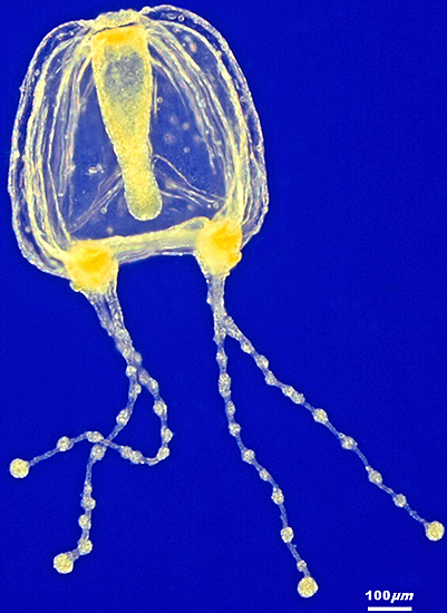 Medusa using Rheinberg illumination [by Carel Sartory]
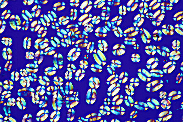 Starch grains using crossed polarisers (20× objective)
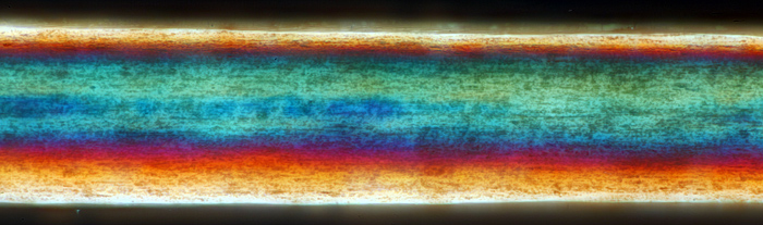 Human hair using crossed polarisers (40× objective)
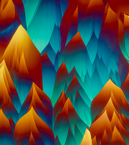 Crystals using crossed polarisers and retarder [by Bevil Templeton-Smith]
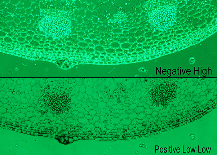 Sunflower stem section using two types of phase contrast (with green filter)
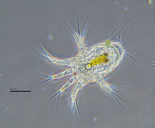 Nauplius larva using phase contrast [by Stephen Durr]
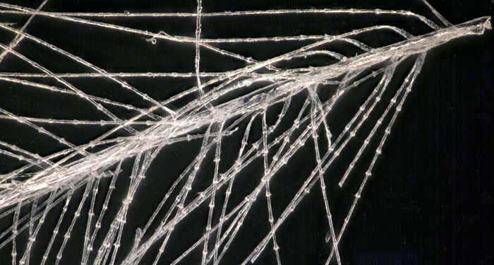
Pam Hamer brought some slides of fibres from electrostatic dusters that we could admire using a small Lomo microscope fitted with a simple polariser and analyser. She also brought prints of photomicrographs of duster fibres and compostable and non-compostable teabags.
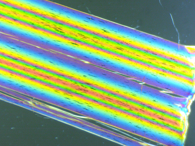 Fibre from electrostatic duster using crossed polarisers (10× objective) [by Pam Hamer]
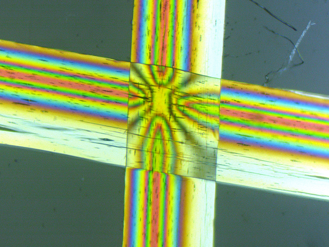 Fibre from electrostatic duster using crossed polarisers (10× objective) [by Pam Hamer]
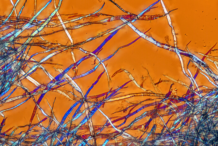
 Non-compostable teabag using crossed polarisers (10× objective) [by Pam Hamer]
Alan Wood brought some prints and notes to illustrate phase contrast, dark-ground and crossed polarisers plus retarders. He also brought equipment for phase contrast and dark-ground.
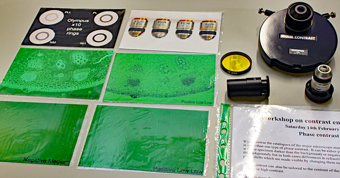 Phase contrast
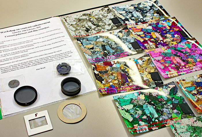 Crossed polarisers and retarders
Alan brought some equipment for dark-ground that is very expensive if bought from a major microscope manufacturer. He also showed how he uses a set of 35 mm dark-ground stops from eBay with an Olympus Abbe condenser that does not have a filter tray.
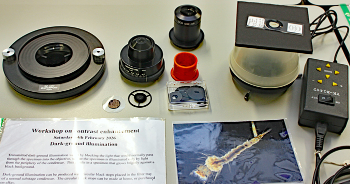 Dark-ground
He also showed how to produce dark-ground illumination for a stereo microscopes using an inverted-ring-light and a pudding basin with its base removed. It raises the specimen above the stage, so needs a stereo microscope with either a tall stand or a 1.5× supplementary objective
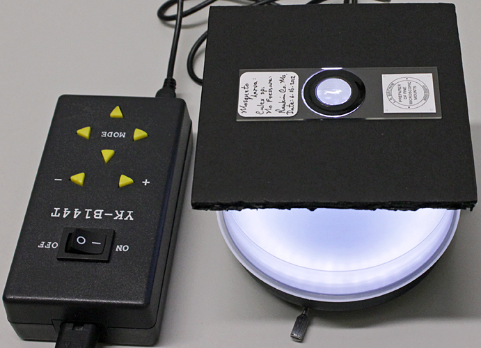 Dark-ground illuminator for a stereo microscope
 Mosquito larva using dark-ground illumination
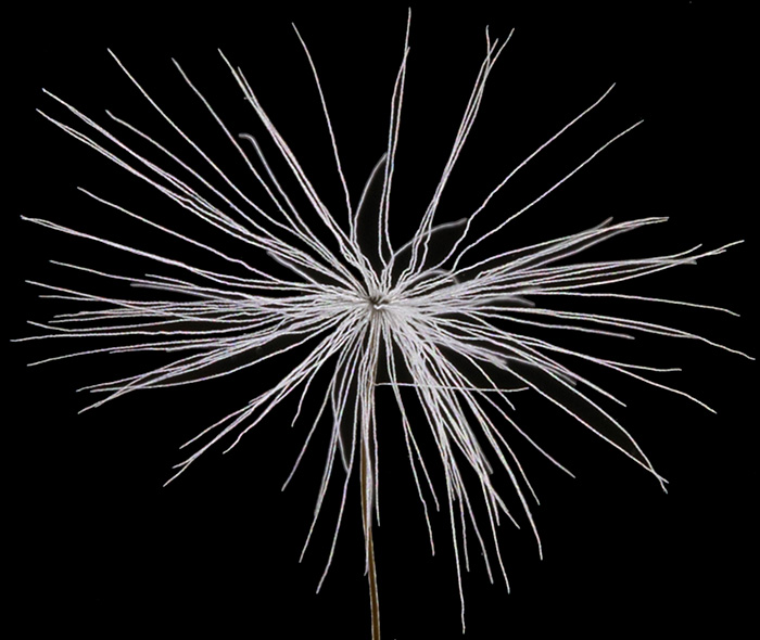 Dandelion seed using dark-ground illumination
Photomicrography
Inevitably, the subject of cameras for use with microscopes was raised. For use via USB with a computer, the simplest are probably eyepiece cameras that fit into a standard 23.2 mm eyepiece tube, or fit stereo microscopes that have wider eyepieces using adapters that are often included.
Chris showed us his 5 MB Hayear C-mount camera that he uses in conjunction with a 0.5× reducing lens to shrink the image down to the size of the small sensor.
Acknowledgements
Our thanks to the Natural History Museum for allowing us to use the Angela Marmont Centre and their Olympus CX41 and CX43 binocular compound microscopes.
Report and most photographs by Alan Wood