

Down House Microscopy Weekend
Saturday 16th and Sunday 17th August 2025
This was our second visit to Down House in Kent, where Charles Darwin lived for 40 years. Quekett member Tim Fullick is one of the volunteers at Down House, and in the Discovery Suite he set up a display of brass microscopes similar to the ones that Darwin used.
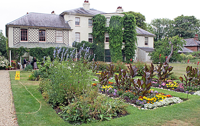 Down House
This year, we had additional displays in the garden laboratory that is attached to the greenhouse and was built for Darwin. With more space (and no fragile antiques), these extra displays were designed to be hands-on for families.
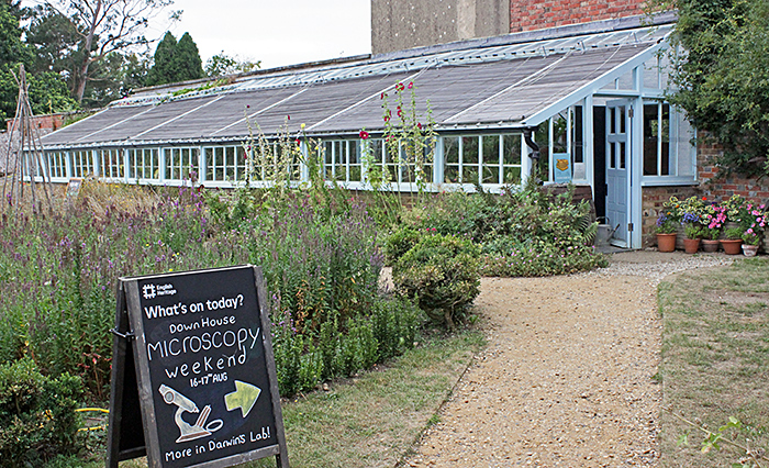 Greenhouse
Discovery Suite
Upstairs in Down House, we had displays of antique microscopes, carnivorous plants, orchids, seeds and aquatic life.
Tim Fullick showed four antique brass microscopes, similar to the ones that Darwin used at various points in his career.
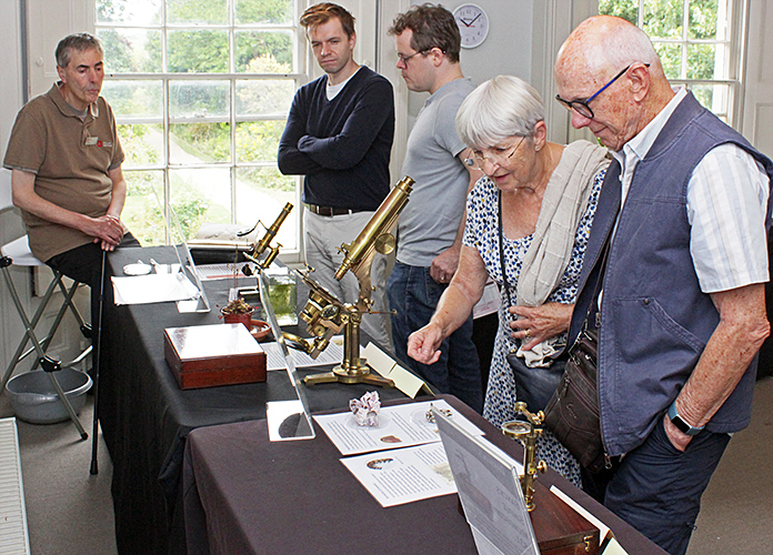 Tim Fullick (left) with visitors
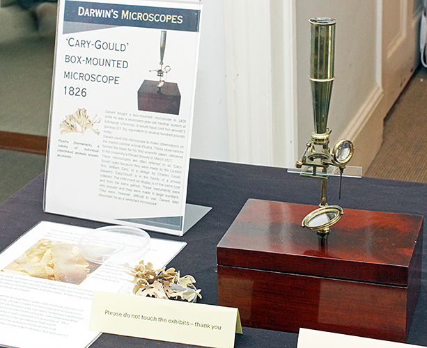 Cary-Gould box-mounted microscope
While Darwin was a student, he used this type of microscope to examine hornwrack, a colonial animal that was then known as Flustra carbasea.
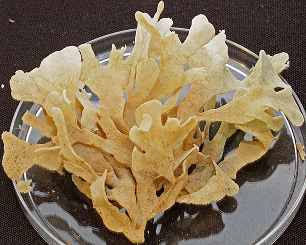 Bryozoan (Flustra carbasea, now called Carbasea carbasea)
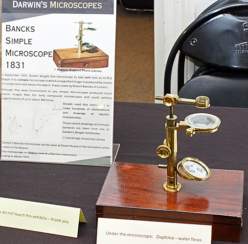 Bancks Simple microscope
Darwin purchased a simple microscope like this one in 1831, to take with him on H.M.S. Beagle. He used it to make hundreds of observations and drawings of aquatic invertebrates. Tim set it up to observe live Daphnia in a watchglass. The microscope was in very poor condition when Tim acquired it, but it has since been expertly restored by Quekett member Penny Thoyts.
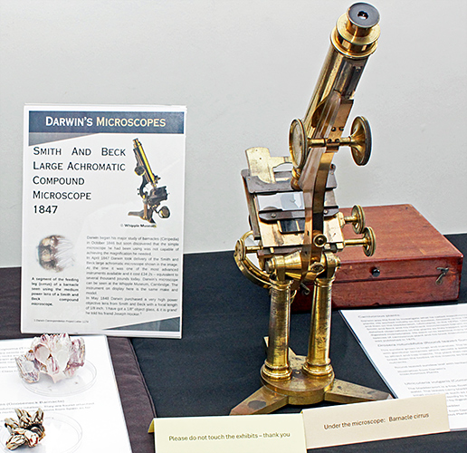 Smith & Beck Large Achromatic microscope
Darwin purchased an advanced microscope like this one in 1847, to help him study barnacles (Cirripedia). Tim used the microscope to show a slide of a barnacle cirrus, and brought examples of titan barnacles (Megabalanus coccopoma) and gooseneck barnacles (Pollicipes pollicipes)
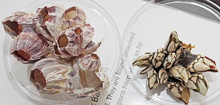 Barnacles
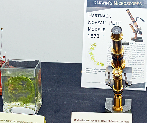 Hartnack Nouveau Petit microscope
Darwin purchased one of these microscopes in 1873. Tim used it to show a slide of the head of a Drosera tentacle. Tim brought a potted round-leaved sundew (Drosera rotundifolia) and a small tank with the aquatic common bladderwort (Utricularia vulgaris). He had added some Daphnia to the tank, and many of them had been trapped in the bladders.
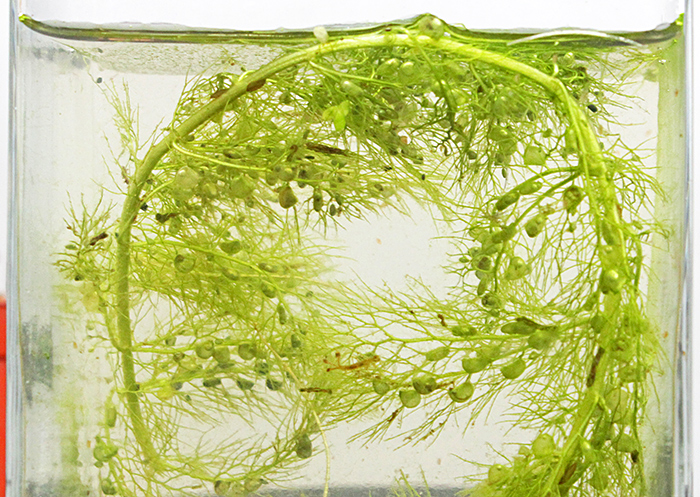 Common bladderwort and waterfleas
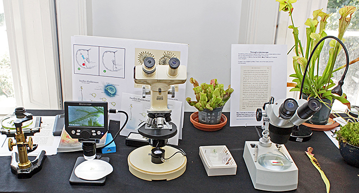
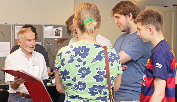
Joan Bingley brought some prepared slides and used a range of microscopes to show various aspects of carnivorous plants (bladderworts, pitcher plants and sundews).
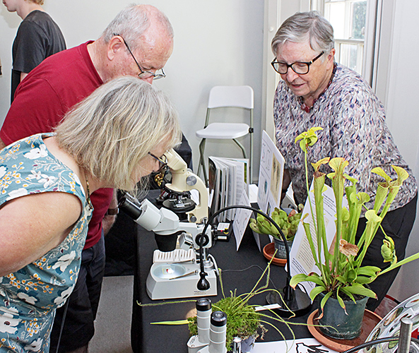 Joan Bingley (right) with visitors
A small digital microscope was set up to show bladders of Utricularia vulgaris with trapped waterfleas (Daphnia), visible because of their black eyes.
Bladderworts pump water out of their bladders to distort the walls. There is a trap door to keep the bladder closed until a small creature touches a trigger hair that opens the trap door, releasing the walls and sucking the prey into the bladder. For the full details, see this paper: The biomechanics of fast prey capture in aquatic bladderworts.
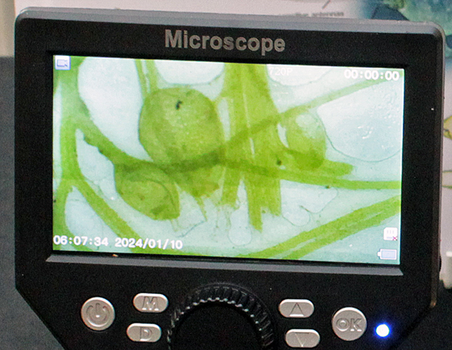 Daphnia trapped by common bladderwort
The microscopes included a simple microscope on a focusing stand, a Koolertron digital microscope with built-in screen, an ex-laboratory Wild M11 compound microscope converted to LED illumination, and a small Brunel stereo microscope.
 Carnivorous plants
Sundews have sticky leaves that trap and slowly digest insects. The one on display (Drosera filiformis) has long, thin leaves, and with the aid of a magnifying glass we could see the tiny insects that it had trapped.
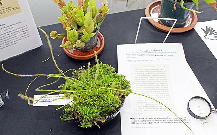 Sundew (Drosera filiformis) and pitcher plant
The pitcher of pitcher plants has a smooth rim so that the insects that it lures with nectar slide down into the digestive fluid. The inside of the pitcher is waxy or has downward-pointing hairs, so that it is very difficult for insects to climb back up and escape. One of the volunteers sliced open a pitcher so that we could see how many insects had become trapped and were being slowly digested.
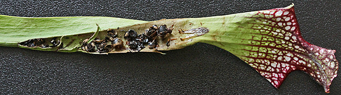 Insects trapped in pitcher plant
Terry Hope focussed on two aspects of pollination in orchids. Darwin was sent specimens of the epiphytic orchid Angraecum sesquipedale from Madagascar, which has nectar spurs around 33 cm long, and he surmised that it was pollinated by a moth with a very long proboscis. The pollinator was eventually found to be a hawkmoth and was named Xanthopan morganii ssp. praedicta. Darwin observed that when the pollinating orchid bees (Euglossini) touched a seta in the flower of Catasetum saccatum, the pollinia (which contains sacs of pollen) was forcibly ejected onto the bee. He was unable to discover the mechanism.
 Terry Hope (left) with visitors
Terry used a Chinese inspection camera to display a large image of an orchid flower on a monitor. There were a few whiteflies in the flower, which attracted a lot of attention.
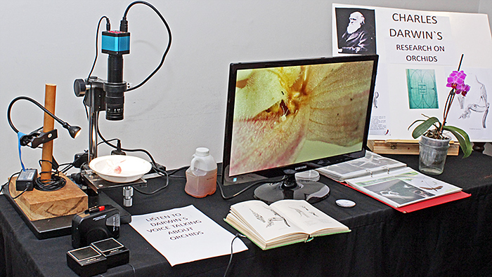 Terry Hope’s display
Terry also showed a copy of Darwin’s book On the Various Contrivances by Which British and Foreign Orchids Are Fertilised by Insects, and On the Good Effects of Intercrossing, and had an audio recording of himself reading an extract from the book.
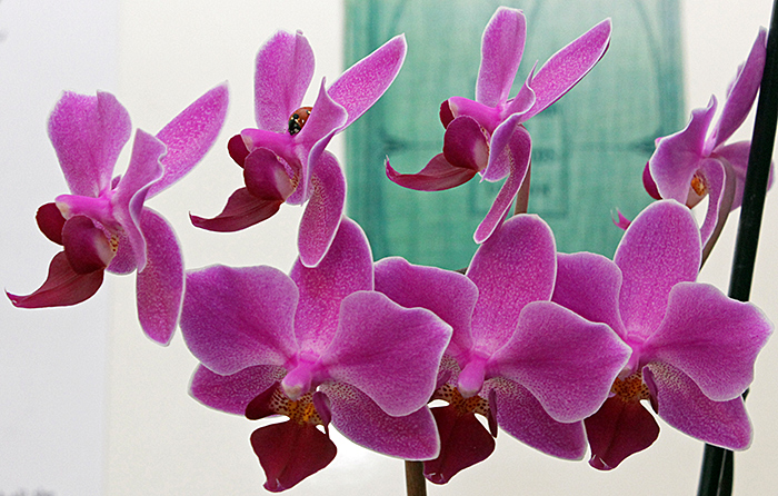 Potted orchid in full bloom
Lisa Ashby put on a display about seeds, explaining the wide range of sizes and the various methods of dispersal. She provided some small microscopes for a closer look at some seeds.
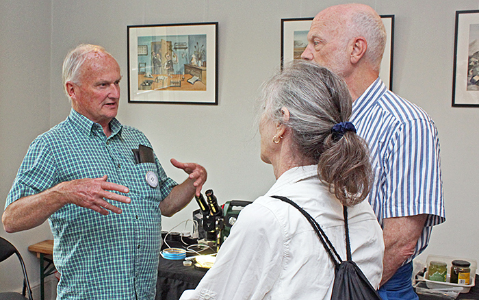
 Lisa Ashby with visitors
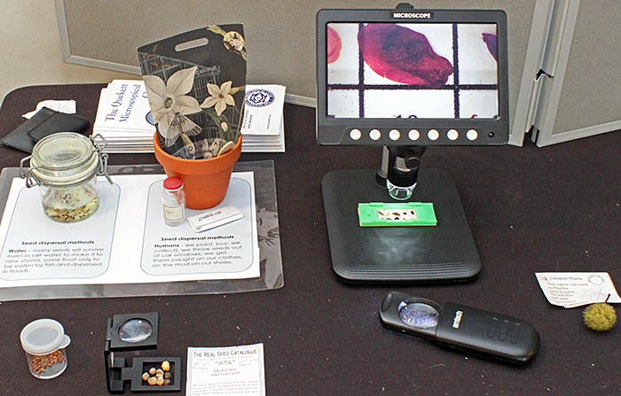 Lisa Ashby’s display
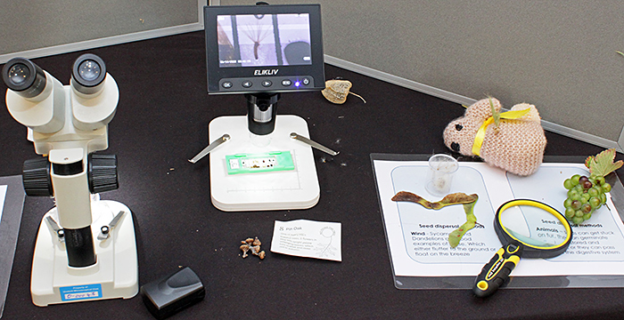 Lisa Ashby’s display
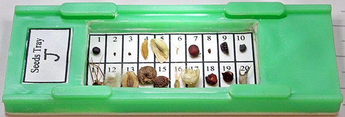 Slide of dry-mounted seeds
Slides like this are sold for mounting microfossils or foraminifera, but they are obviously good for seeds too.
Mark Berry brought five microscopes that would have been used in laboratories, and that are still used by enthusiasts. They were a Watson Microsystem 70 compound microscope in near-mint condition, a Gillett & Sibert Conference compound microscope, a Zeiss Standard inverted microscope, and stereo microscopes by CTS and Zeiss Jena. He had converted most of them to use LED illumination.
 Mark Berry with visitors
Mark used his microscopes to show live flatworms (Dugesia sp., Turbellaria) from fresh water, live marine colonial ciliates (Carchesium ?), live marine bryozoans (Bowerbankia sp.), three species of lichen, and smears of his own blood that he prepared and stained on the day.
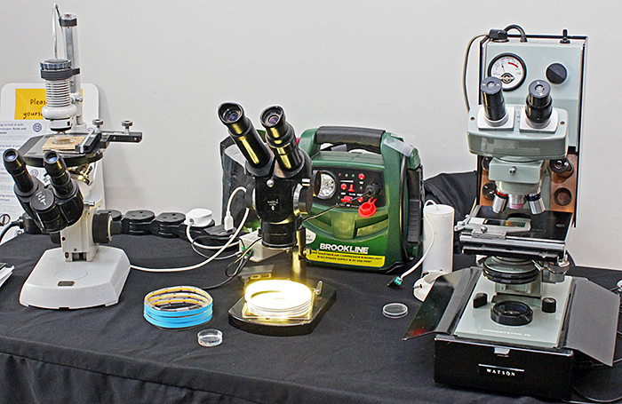 Mark Berry’s display
Garden Laboratory
The permanent attraction in the laboratory is an observation beehive. It is much taller and narrower than a normal hive, with glass sides and only a few frames. There is a clear tube through the wall of the laboratory, so that visitors can watch the bees coming and going, as well as watching their activities inside the hive. We provided magnifying glasses to give visitors a closer look at the bees. Our displays included bee-related items such as slides of pollen and bee parts, and a guide to recognising the Asian hornet.
 Observation beehive
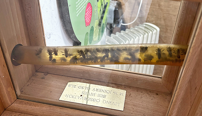 Entrance to beehive [by Pam Hamer]
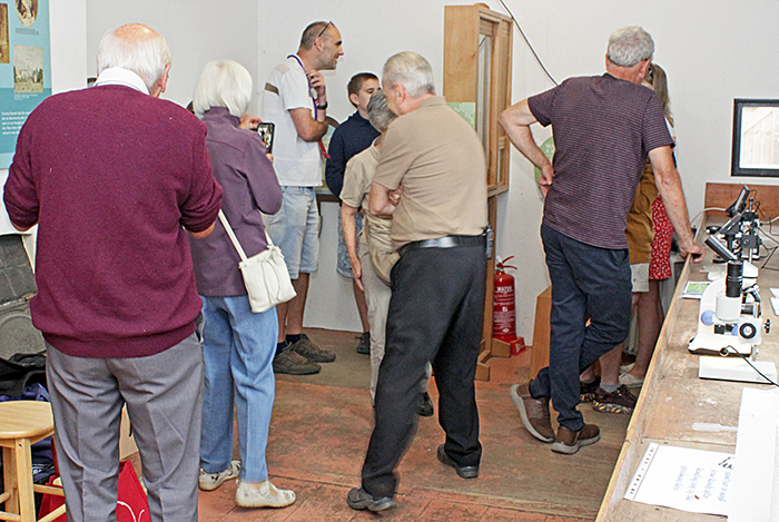
Pam Hamer and Martin Parnham showed a variety of small optical and digital microscopes that are suitable for children. Their specimens included minerals, fossils, live woodlice, plant parts from the garden, galls, and slides of crystals, pollen, small insects (including an aphid, a head louse and Drosophila) and parts of honeybees. They also had an Asian hornet (Vespa velutina) encased in resin, the one that kills large numbers of honeybees.
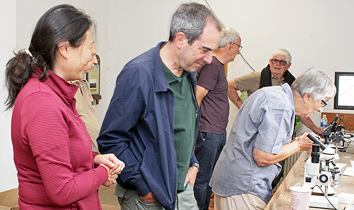 Martin Parnham, Pam Hamer and visitors
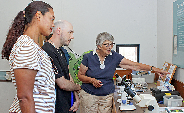 Pam Hamer with visitors
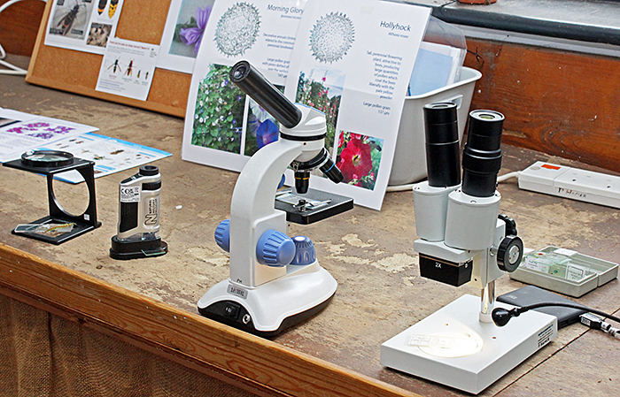 Small optical microscopes
 Digital microscopes
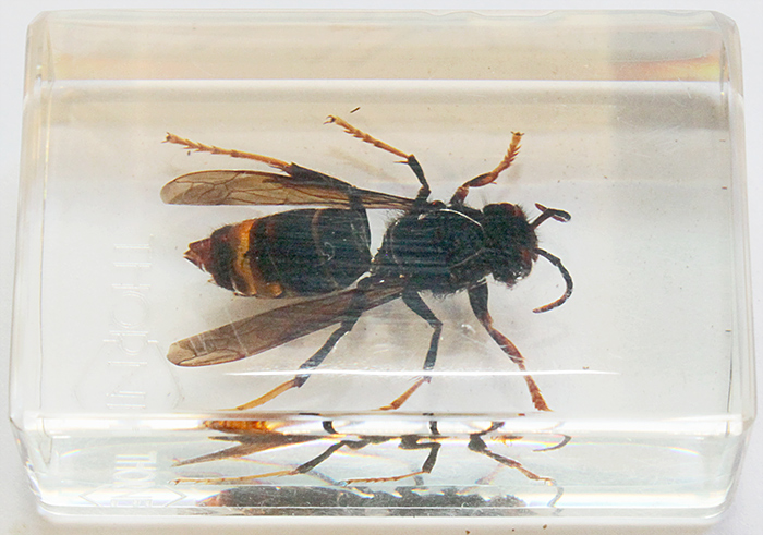 Asian hornet
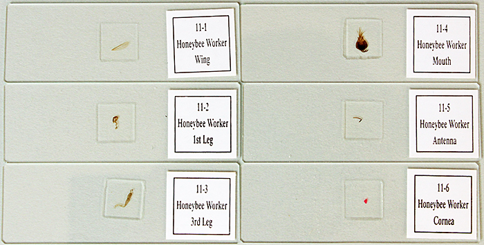 Slides of honeybee parts
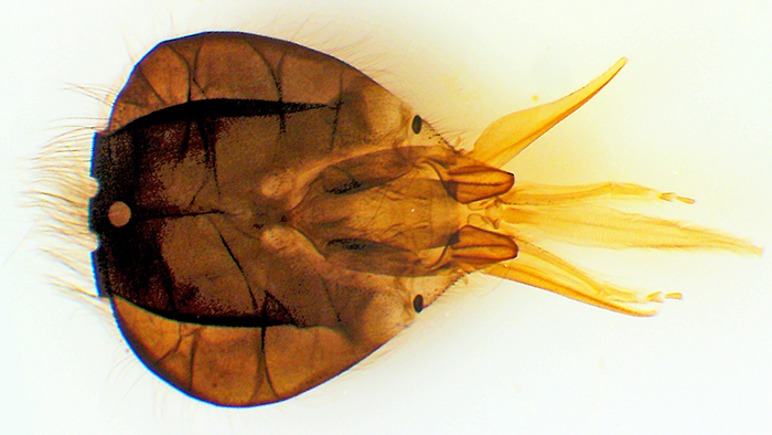 Head of honeybee [by Pam Hamer]
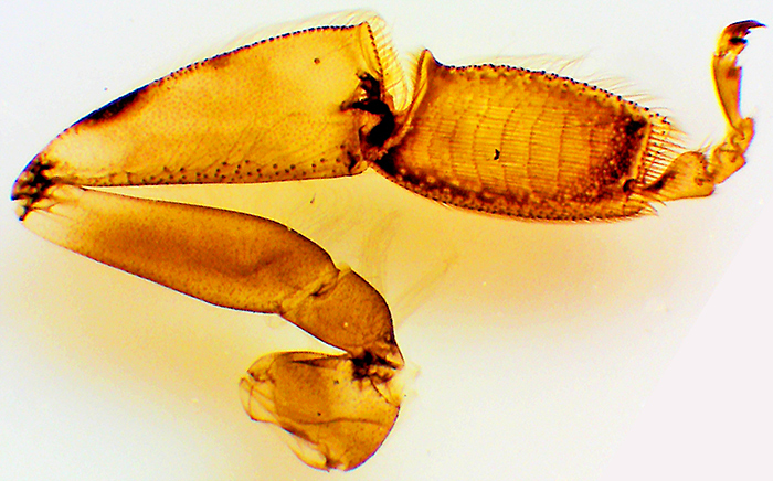 Hind leg of honeybee [by Pam Hamer]
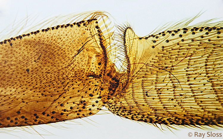 Pollen basket on hind leg of honeybee [by Ray Sloss]
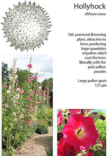 Hollyhock poster
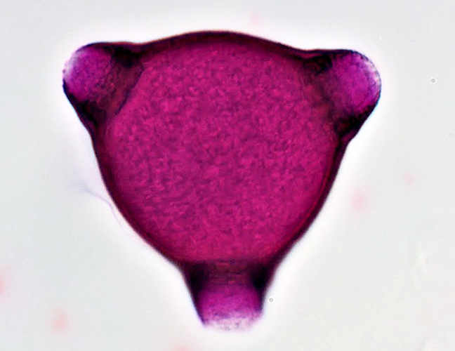
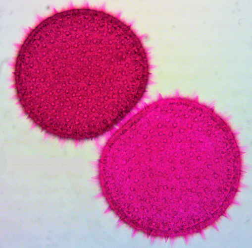 Hollyhock pollen [by Pam Hamer]
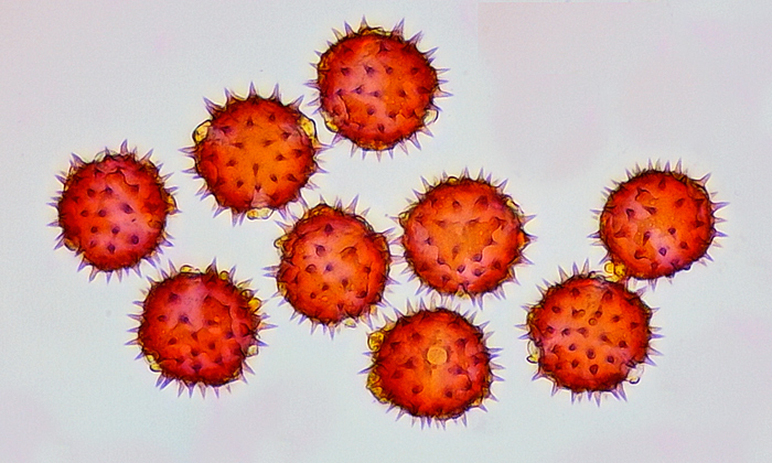 Sunflower pollen [by Gordon Brown]
 Rosebay willowherb pollen [by David Galliford]
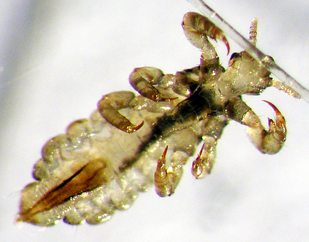 Head louse (Pediculus humanus capitis) [by Chris Thomas]
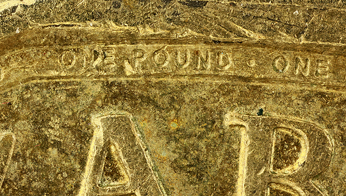 Micro writing on a £1 coin
Chris Thomas brought two monocular microscopes from the 1960s, both brought up-to-date with LED illumination. One of them was fitted with a digital C-mount camera instead of an eyepiece, so that images could be seen and captured with ToupView software on a laptop computer. Chris not only showed visitors slides of pollen and leaf prints, he guided them through making their own slides.
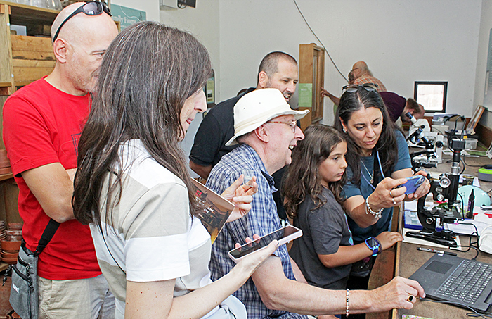 Chris Thomas (centre) with visitors
It is not easy to observe features such as stomata on the surface of leaves, and traditionally clear nail varnish has been used to make an impression that can be viewed under a microscope, but now there is a better technique. Chris provided fresh leaves of Portuguese laurel, ivy and Iris, and demonstrated how to cut a square (15–20 mm) from a leaf and press it onto a drop of Vida Rosa UV resin on a slide. He then covered the leaf with non-stick baking paper, placed another slide on top, and clamped the sandwich with two small metal clips. Then he put the assembly upside down (resin nearest the bulbs) into a UV lamp that is designed for setting nail polish, and exposed it for 60 seconds. This hardens and dries the resin. After separating the parts and peeling off the leaf, the slide is ready to be labelled and examined under a microscope.
Chris guided visitors through the process, so that they could make a leaf peel slide and take it home.
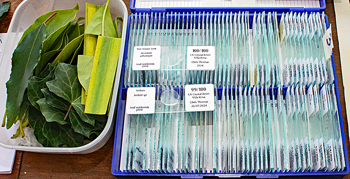 Slides of leaf peels
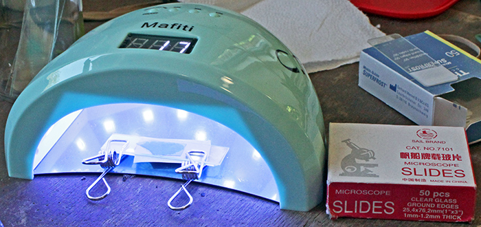 Curing resin with UV nail polish lamp
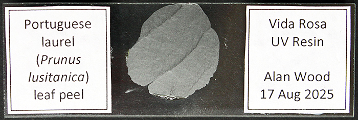 Leaf peel of Portuguese laurel
It is easy to design slide labels like this in Microsoft Word, using borders on cells in a table.
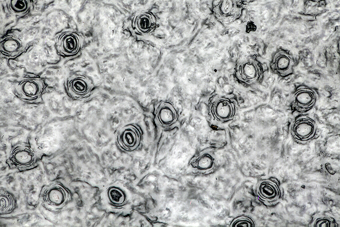 Leaf peel of Portuguese laurel (40× objective)
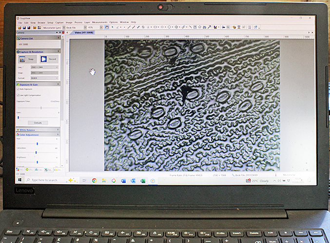 Leaf peel under a microscope, viewed on a computer
Chris also brought a box of his slides of stained pollen, so that we could examine them under a microscope.
 Slides of pollen
Chris also showed visitors and volunteers how to use a smartphone adapter to take photographs through a microscope (photomicrographs). The adapter clamps onto eyepieces of various diameters, and has a platform covered in suckers that hold the phone with its camera lens centred over the microscope eyepiece.
 Chris Thomas explaining the smartphone adapter
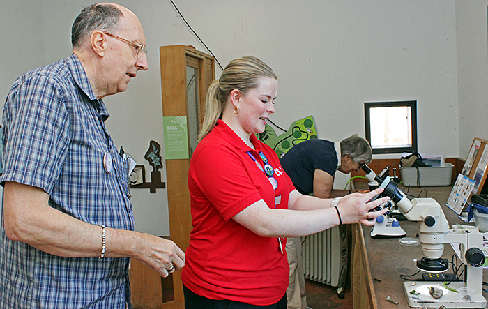
Alan Wood brought his ex-laboratory Olympus SZ4045 zoom stereo microscope from the 1980s, given a new lease of life with an LED ring-light. He used it to show specimens from the gardens and the greenhouse.
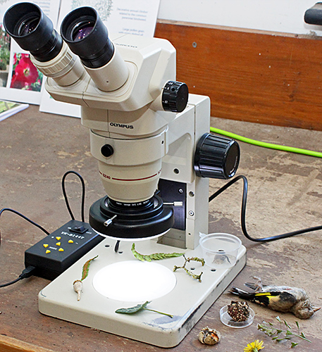 Stereo microscope
His specimens included flowers, fern sori, hollyhock seeds, poppy seed heads, lichen on small twigs, and a small mouldy apple. He also showed a dead goldfinch that was dropped by a small raptor that accidentally flew into the laboratory.
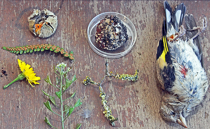 Some of Alan Wood’s specimens
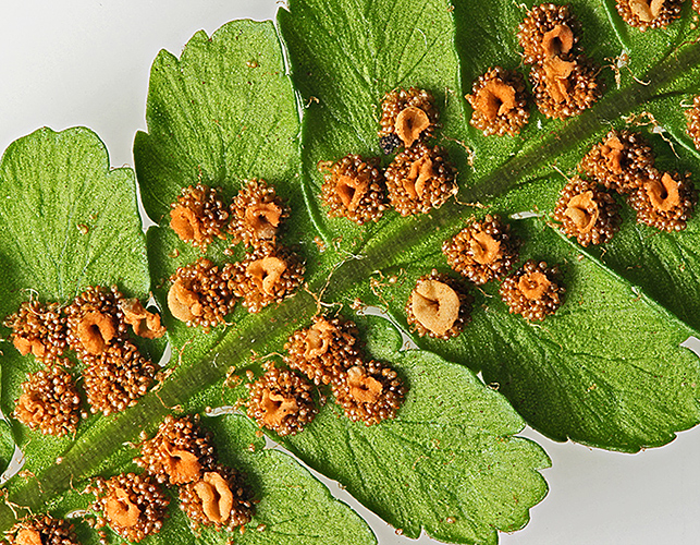 Sori on underside of a fern frond
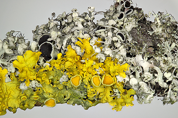 Lichen on a twig
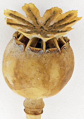 Seed head of a poppy
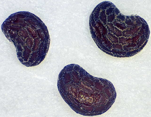 Poppy seeds (0.75 mm)
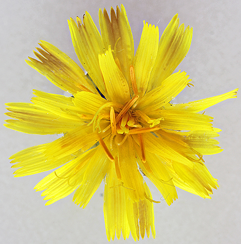 Hawkbit flower
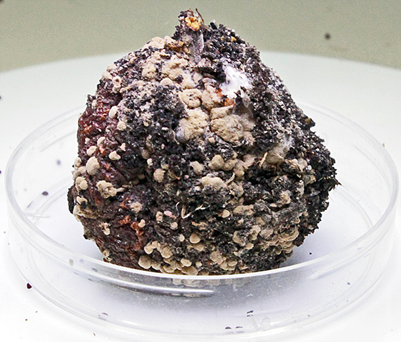 Small mouldy apple
The plants in the greenhouse include carnivorous plants, orchids and ferns.
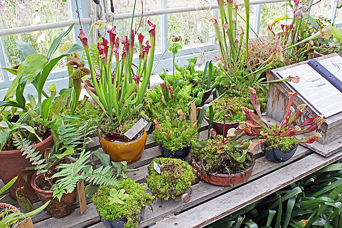 Greenhouse plants
Some of the flower pots are in saucers where algae are growing. Alan used a pipette to take some samples into the laboratory, and found some rotifers among the algae.
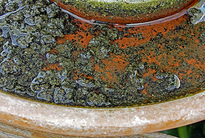 Algae in flower-pot saucer
English Heritage recorded 488 visitors to Down House over the weekend. Massive thanks to the English Heritage volunteers, organised by Moira McManus. They marshalled the visitors, so we were not swamped, while encouraging them to see and interact with all our displays in the Discovery Suite and the laboratory. We are planning to repeat the event next year, on Saturday 22nd and Sunday 23rd August 2026.
Report and most photographs by Alan Wood