

Quekex25 – The Annual Exhibition of Microscopy
Saturday 11th October 2025
The Club’s Annual Exhibition of Microscopy (Quekex25), was held for the fifth time at Elm Court Youth & Community Centre, Potters Bar, Hertfordshire. There were demonstrations and exhibits by members, displays of the photomicrographs, videos, slides and artwork submitted for awards, a lecture, and plenty of gossip.
Quekett members can view galleries of the photomicrographs and videos submitted for Barnard Awards, slides submitted for Eric Marson awards, and artwork here:
• Quekex25 Galleries
Exhibits
This year, the theme for exhibits and demonstrations by members was “I made it or I modified it”.
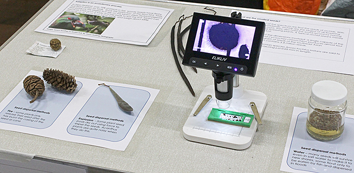
Lisa Ashby showed her display about seeds from the Down House Microscopy Weekend, explaining the wide range of sizes and the various methods of dispersal. She provided some small microscopes for a closer look at some small seeds.
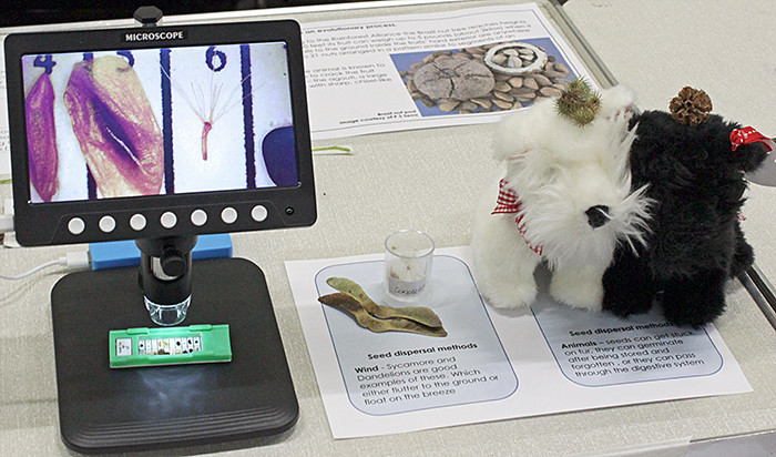 Lisa Ashby’s seed exhibit
 Lisa Ashby’s seed exhibit
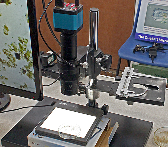
Nigel Ashby showed the latest version of his apparatus for showing specimens larger than normal slides. The stand is from a Watson Greenhough, with a small light-box under a floating stage from a Watson Zoom. Above the specimen are a concave lens, a plumbing pipe, a cubical Chinese inspection camera and a screen from a security camera.
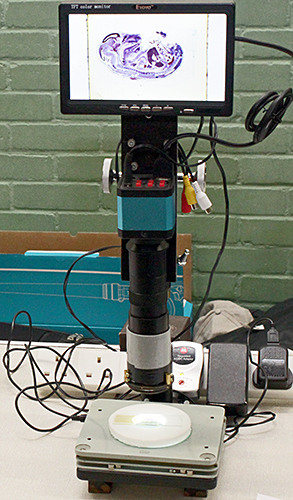 Nigel Ashby’s exhibit
Elizabeth Beston runs the Plankton Project which collects information about marine plankton around the coast of Norfolk, and this was her first time at Quekex.
 Elizabeth Beston with visitors
Elizabeth used small Brunel stereo and compound microscopes to show live plankton samples, and used a GXCAM Hi-Chrome-HR4 camera (purchased with a grant from the Quekett) to show images on a monitor.
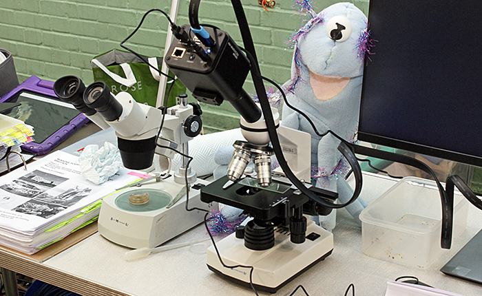 Elizabeth Beston’s exhibit
Joan Bingley brought an assortment of everyday items that she uses in microscopy or bryology, including a chopping board, an eye-dropper bottle, a tea strainer, a Pringles tube (for storing small Petri dishes), acupuncture needles, black and white tiles and a spray bottle. Less usual items were a paint scraper for sampling tree bark substrates for pH tests, and a clinometer to measure angles of substrates in the field.
 Joan Bingley’s exhibit
Relevant to her lecture in the afternoon, Joan also brought some books about mosses, some slides of moss made by Quekett members, and small stereo and digital microscopes for viewing the slides.
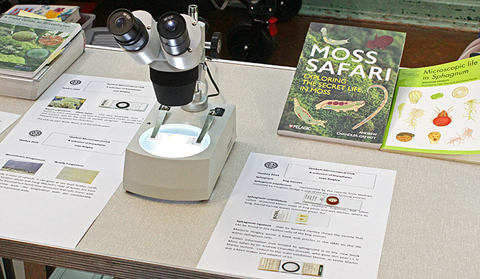 Joan Bingley’s exhibit
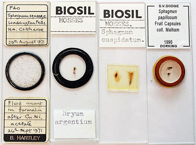 Joan Bingley’s slides
The books that Joan brought were Mosses and Liverworts of Britain and Ireland: A Field Guide by I. Atherton, S. Bosanquet & M. Lawley, A Field Guide to Bryophytes by Dominic Price & Clive Bealey, Microscopic life in Sphagnum by Marjorie Hingley, Moss Safari by Andrew Chandler-Grevatt, and The hidden world of mosses by Neil Bell.
Gordon Brown brought the automated slide stainer that he had previously shown on Zoom meetings. It was not working when it was given to him, but he soon replaced the motor and got it working. Slides are dipped into a series of containers for a set time, so timings can be adjusted by using two or more containers with the same liquid. Gordon now has a protocol for his modified Wacker stain.
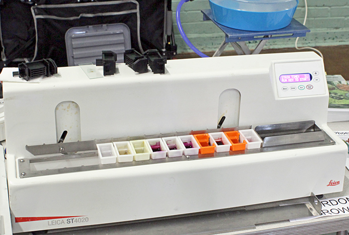 Gordon Brown’s Leica ST4020 Linear Stainer
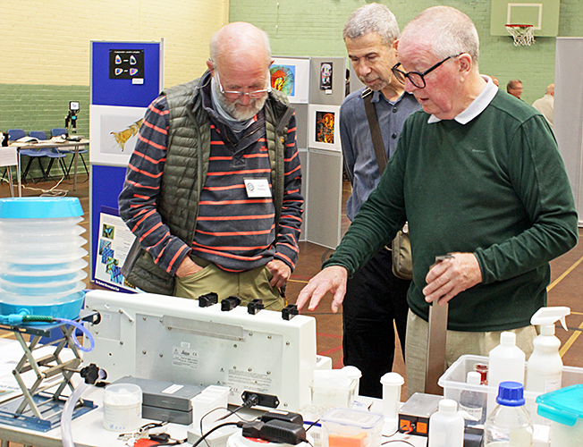 Geoff Phillips, Claudi Soler and Gordon Brown (right)
Gordon also brought Olympus BHTU and BHSP microscopes in which he had replaced the electronics while keeping the voltage indicators working, and showed us the LEDs with their heat sinks.
 Gordon Brown’s LED for Olympus BHS
 Gordon Brown’s LED for Olympus BHT and BHTU
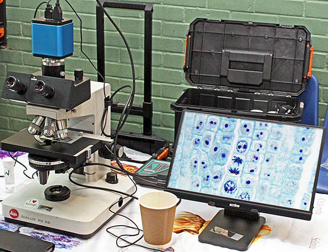
Steve Durr used his trinocular Leitz Dialux 20 EB with Fluotar objectives to show slides of mitosis in Lilium anthers and Allium root apex. He used a camera to display the images on an Aztine monitor.
 Steve Durr’s exhibit
Phil Greaves makes slides of amber with fossil inclusions by cutting the pieces to a suitable size, grinding and polishing, and then mounting in a cell on a slide. He builds up layers of Canada balsam before adding a cover slip. The process produces mounts that are suitable for up to about 200× magnification.
 Phil’s amber slides
 Phil’s amber slides
Phil also brought several items that were made by other Club members. Leslie Martin, Tony Saunders-Davies and Geoffrey Fryer made various styles of compressoria:
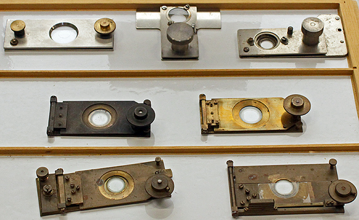 Compressoria
Ainslie made a range of lenses to be inserted above objectives for tube length correction (below left). Dick Speight made apertometers for measuring numerical aperture (below right).
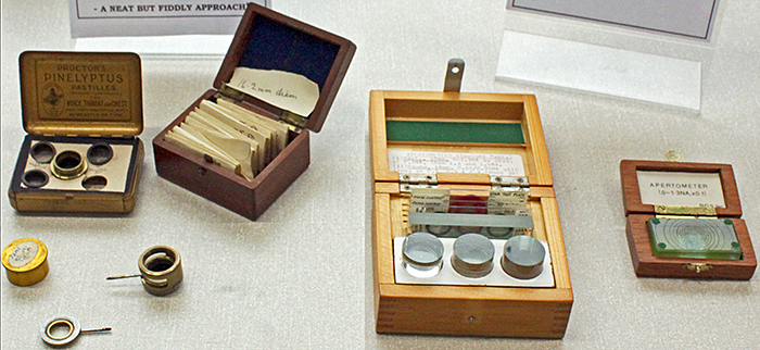 Tube length correction lenses by Ainslie, and apertometers by Dick Speight.
Past president Harry Dade made slides of fungi and fungal spores, and Tony Dutton made slides of large objects such as a phono cartridge and a video sensor.
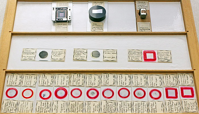 Slides by Tony Dutton (top) and Harry Dade
Pam Hamer showed a novel way of getting children interested in microfossils. The Works sells bags of plastic keyrings that are designed to hold a photograph, but Pam realised that the inside could be coated with PVA adhesive and then sprinkled with fossiliferous sand. The sand she used was from one of the cleaned samples that Adrian Brokenshire used to give away at meetings in Langton Matravers.
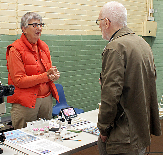 Pam Hamer and Stephen Edler
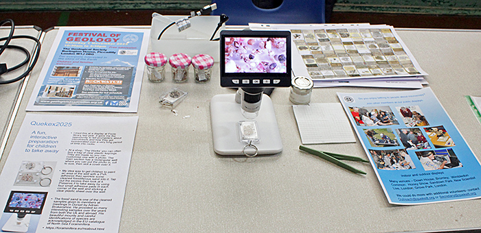 Pam Hamer’s exhibit
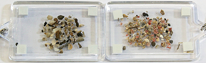 Pam’s sand keyrings
Pam also showed photographs of hundreds of thin rock section slides that have been given to the Club. The slides are smaller than the standard 3×1 inch size, which makes them awkward to store and to use under a microscope.
Terry Hope brought some specimens from his pond and used a Chinese inspection camera to show them on a monitor. The camera was supported on a pole stand, and the specimens were in a small Petri dish on a light box. For viewing specimens on slides, there is a mechanical stage attached to the pole which can be swung out of the way when it is not needed.
 Claudi Soler and Terry Hope (center)
 Terry Hope’s exhibit
Stephen Parker brought three unusual microscopes.
 Danny Ferri and Stephen Parker
Stephen showed two reflected-light microscopes by Leica and Nikon that he had gradually assembled with parts from sales events and eBay. His Nikon Labophot-2 was fitted with three interferometer (DI) 210mm objectives and a long working distance 40× objective. It was fitted with an incident light adapter complete with polariser and analyser, plus an unbranded lamphouse that happened to fit. His trinocular Leitz Laborlux 12 was fitted with a MODULOPAK incident light adapter, complete with polariser, first-order wave plate and iris diaphragm. The objectives were 5×, 10× and 100× BF/DF, 50× BF and 10× interference contrast. His specimens included integrated circuits, forams, mica, calcite and a compact disk.
He also showed a Bausch & Lomb stereo microscope with a stand probably made by Ron Green. The base is from a Vickers M15, with a rotating stage from a Vickers M10a polarising microscope. The stereo head is attached to a tall pole, so that large specimens or a low-power supplementary objective could be used. Stephen has added a polarising film below the stage, and a camera polarising filter below the head, to produce a low-power polarising microscope.
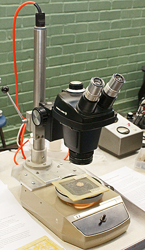 Bausch & Lomb stereo on Vickers base
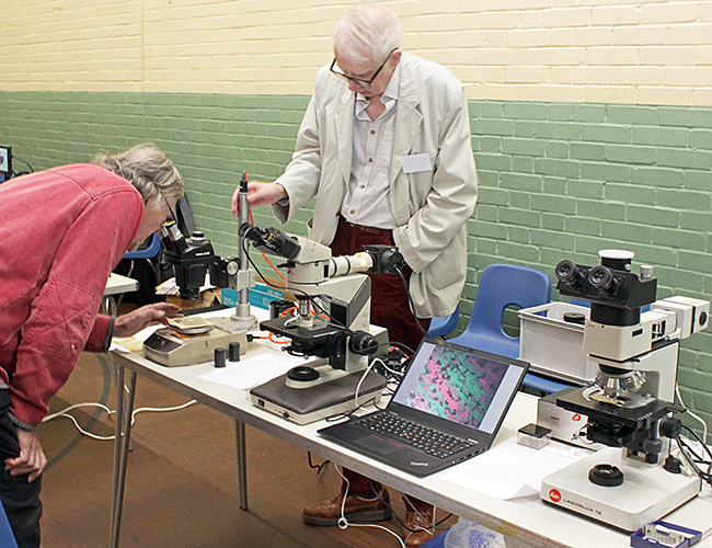
Geoff Phillips normally works with freshwater phytoplankton, but has recently started looking at marine phytoplankton with his Wild M40 inverted microscope. He used a Chinese inspection camera to show images of live plankton on his laptop. He has found that marine coastal waters contain much less phytoplankton than freshwater lakes, and that suspended sediment can be a problem.
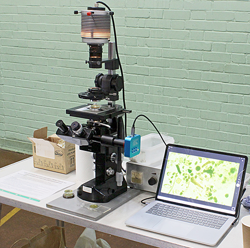 Geoff Phillips’ exhibit
Geoff showed three books, Coastal Plankton: Photo Guide for European Seas by Otto Larink & Wilfried Westheide, Coastal Phytoplankton: Photo Guide for Northern European Seas by Alexandra Kraberg, Marcus Baumann & Clause-Dieter Dürselen, and Marine Plankton: A Practical Guide to Ecology, Methodology, and Taxonomy by Claudia Castellani & Martin Edwards.
Robert Ratford filled three tables with microscopes and specimens, including a trinocular Zeiss Axiostar Plus, a Wild M3X with transmitted-light stand and a Chinese copy of an Olympus SZ4045. His specimens included feathers, rocks, beetle elytra and slides of plant sections.
 Robert’s stereo microscopes
 Robert’s compound microscope
Robert also brought a huge assortment of items that will fascinate fans of the McArthur portable microscopes, including technical drawings, prototype parts, documents and photographs.
 Prototypes for McArthur microscope
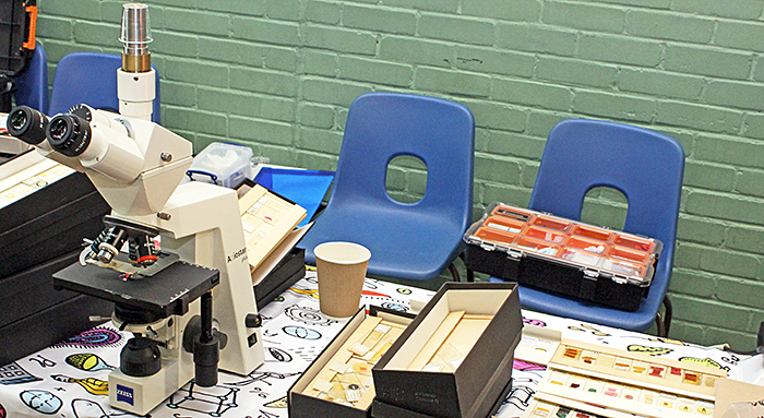
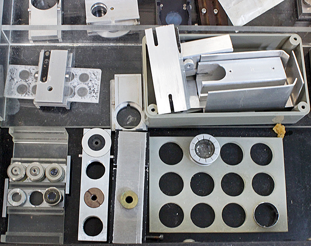
Carel Sartory makes videos of slides of crystals being rotated while viewed under crossed polarisers. Some of his videos were shown on a laptop, and a series of posters explained the equipment that he uses.
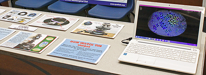 Carel Sartory’s exhibit
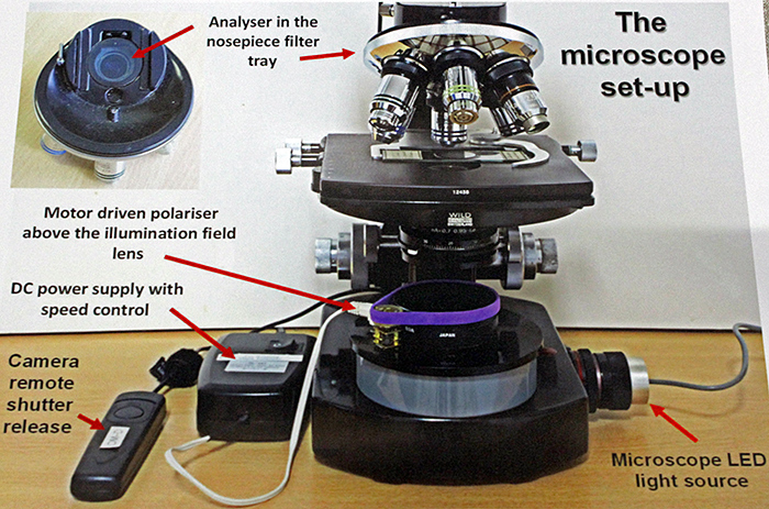 Carel Sartory’s poster
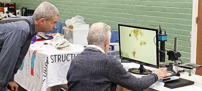
Chris Thomas has made lots of slides of leaf epidermis prints, and has now produced a key for identifying the species. At the Exhibition, Chris was asking volunteers to try out the key, see how well it works, and suggest improvements.
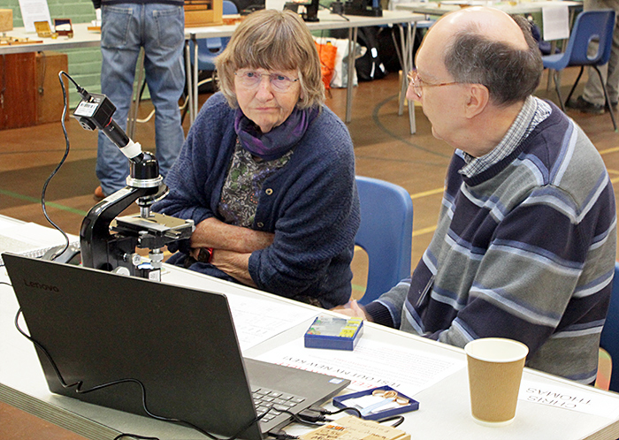 Chris Thomas (right)
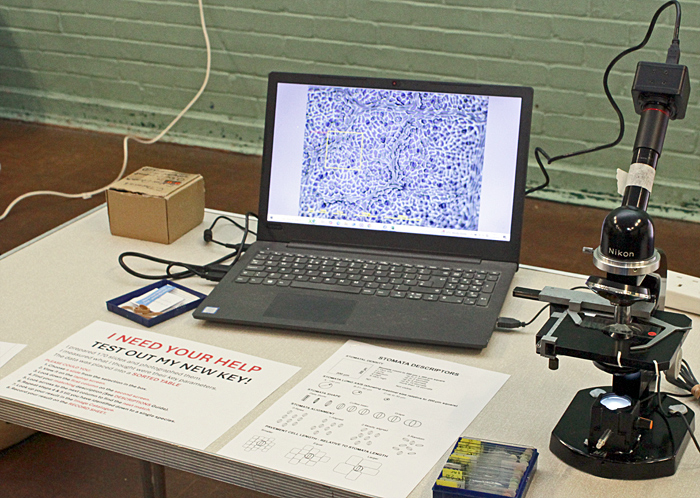 Chris’s exhibit
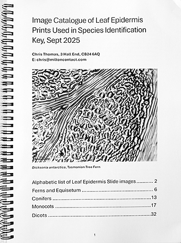 Key to leaf epidermis prints
Nigel Williams used a variety of simple and compound microscopes to show slides made by past presidents:
Peter Sartory: Cribrostomoides
Carel Sartory: Diptera Diopsidae
Phil Greaves: Millipede in Dominican amber
Joan Bingley: Peacock feather
He also showed a slide that he had made, with 33 star-sand forams mounted in Vida Rosa resin.
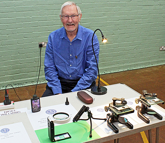 Nigel Williams
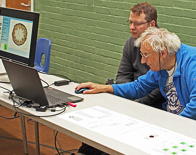
Alan Wood showed how he uses an LED ring-light and a pudding basin to produce shadowless and dark-ground illumination for a stereo microscope. His specimens included a printed circuit, a coin, a key, a watch, batteries, lichen and a slide of a mosquito larva.
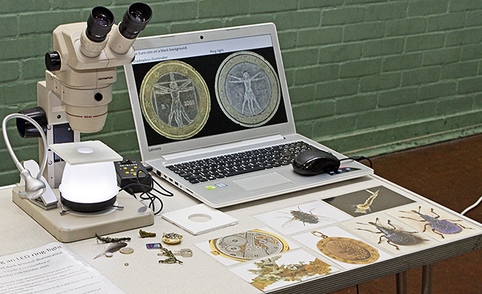 Alan Wood’s exhibit
You can see the slides from Alan’s PowerPoint here:
Click the arrows to move through the slides. Click the symbol at bottom right for a larger version.
Displays
Prints of the photomicrographs that were submitted for Barnard Awards were displayed on panels in the middle of the hall. You can see all the entries on-line in the Quekex25 Galleries.
 Entries for a Barnard Award
 Entries for a Barnard Award
The videos that were submitted for a Barnard Award were shown on a laptop. Charles Hussey was the judge this year.
Graham Matthews was the judge for the Eric Marson Awards for slides, and he showed the actual slides and a PowerPoint presentation of photographs and photomicrographs.
 Luke Whitehouse and Graham Matthews (right)
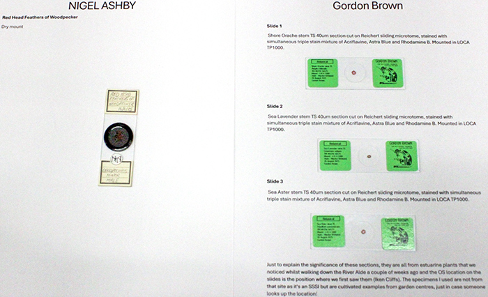 Slides by Nigel Ashby and Gordon Brown
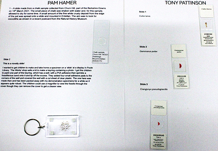 Slides by Pam Hamer and Tony Pattinson
Lecture
The morning lecture was intended to be “Nobel prizes, numbers, sunburns and awe” by Dr Philippe Laissue, but sadly Dr Laissue was unable to attend.
The afternoon lecture was “Adaptability of Bryophytes” by Joan Bingley.
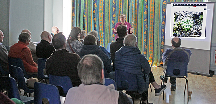 Joan Bingley lecturing
Acknowledgements
Our thanks to everyone who:
- brought exhibits and demonstrations
- submitted photomicrographs, videos, slides and artworks
- judged the photomicrographs, videos, slides and artworks
- booked the venue
- got out and packed away the tables and chairs
- organised the displays of photographs
- organised tea, coffee and biscuits
- arranged, broadcast and recorded the lectures
- publicised the event and the meeting report on social media
We hope to see you all again next year!
Report and most photographs by Alan Wood