

Warnham Local Nature Reserve excursion
Saturday 9th August 2025
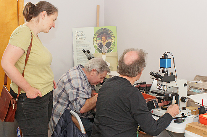
This was the fourteenth Quekett excursion to Warnham Local Nature Reserve on the outskirts of Horsham, West Sussex, hosted by the Friends of Warnham Local Nature Reserve, and organised by Graham Matthews. As in recent years, we set up our microscopes, cameras and computers in a small room at the side of the Discovery Hub.
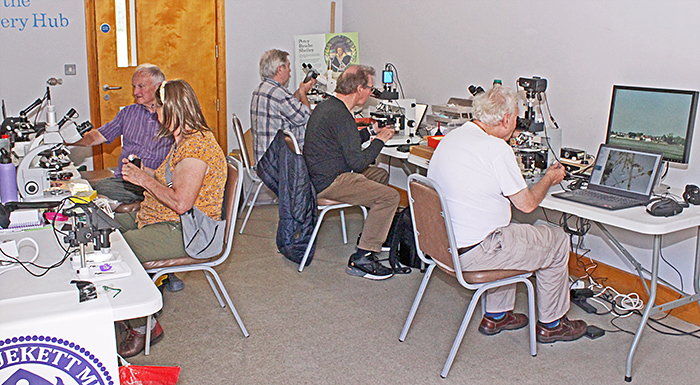 Quekett members in the Discovery Hub
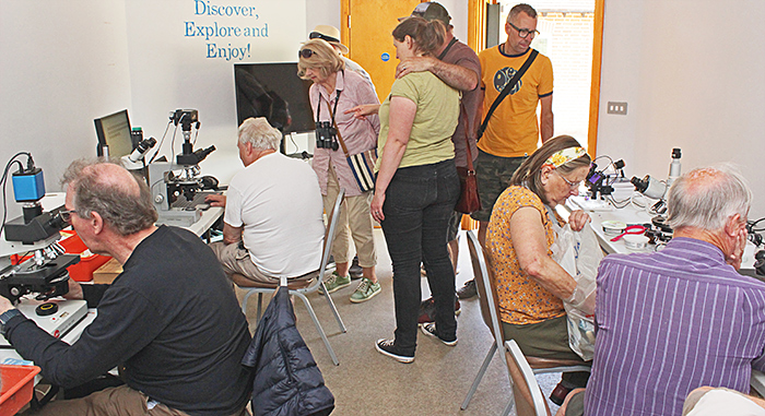 Visitors and Quekett members in the Discovery Hub
The Reserve includes a large millpond, three dipping ponds with boardwalks, a reed bed, the Shelley pond, two streams, meadows and woodlands.
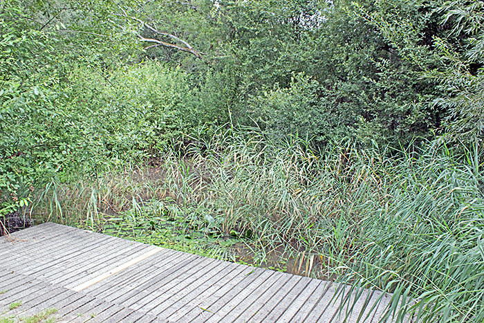 Dipping pond
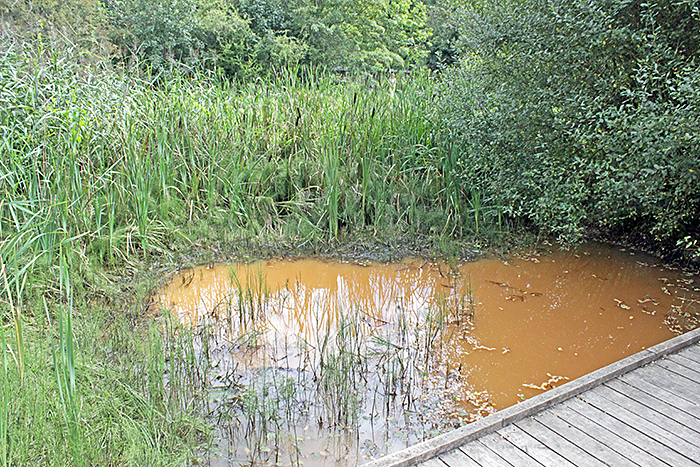 Dipping pond
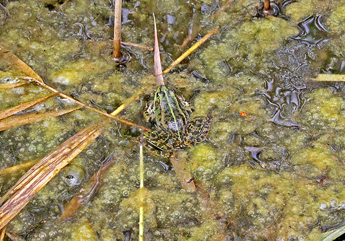 Marsh frog in the reed bed
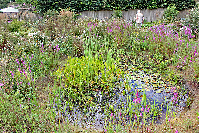 Shelley pond
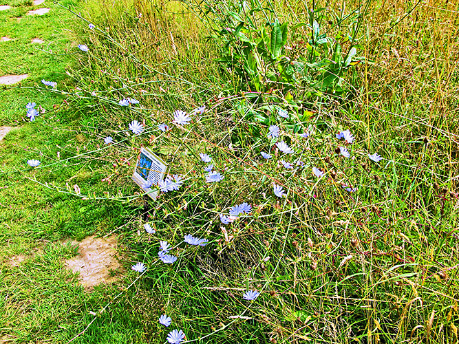 Chicory (Cichorium intybus) in the meadow [by Graham Matthews]
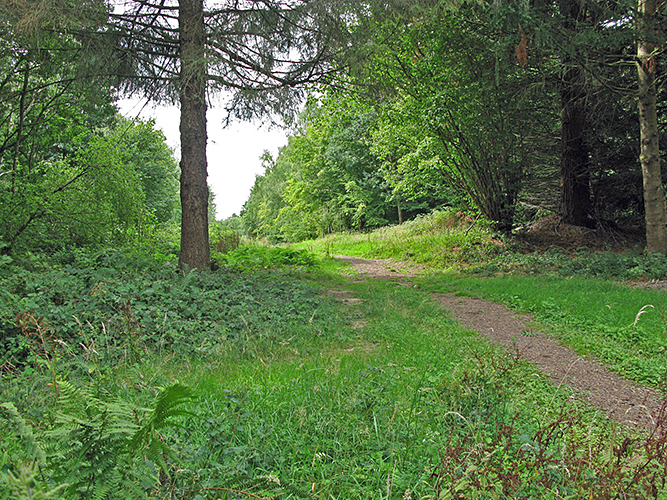 Woodland path [by Graham Matthews]
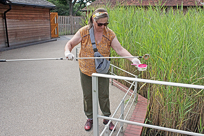
For collecting samples from the pond, we used a plankton net, a small bucket, and small jars on the end of telescopic golf-ball retrievers. The jars were held in place by sticky tape, rubber bands or clamps.
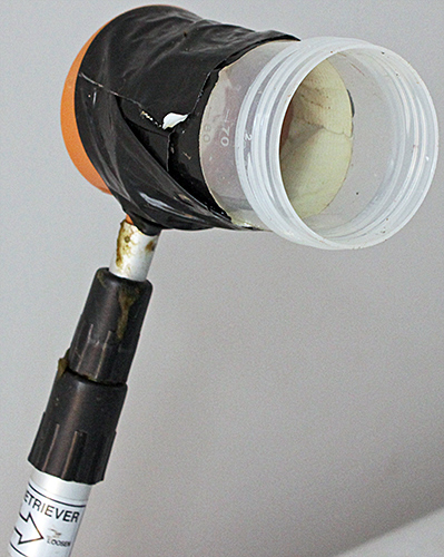 Golf-ball retriever
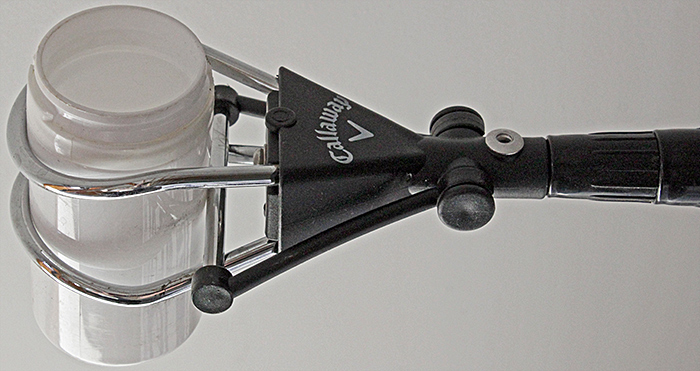 Golf-ball retriever
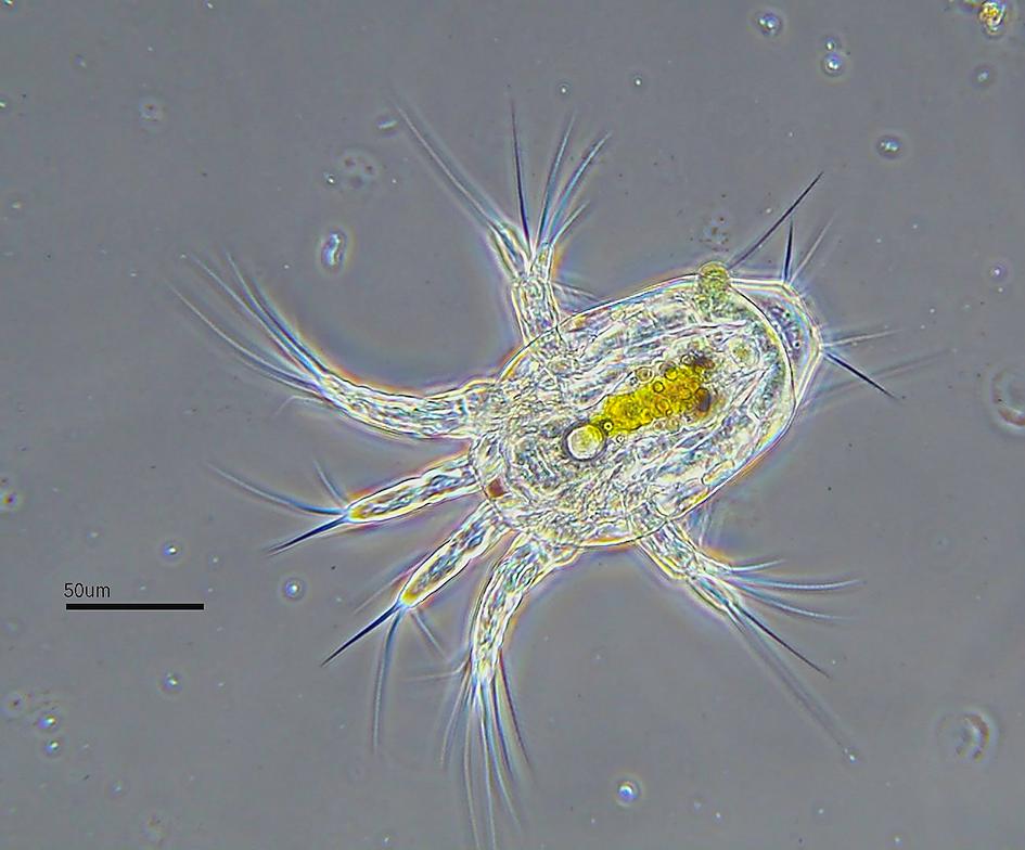
The specimens that we found in the samples included diatoms, desmids, rotifers, ciliates (including peritrichs), copepods, Cladocera, ostracods, mayfly nymphs, mites, gastrotrichs, testate amoebae, a leech, green hydra (Hydra viridissima) and algae.
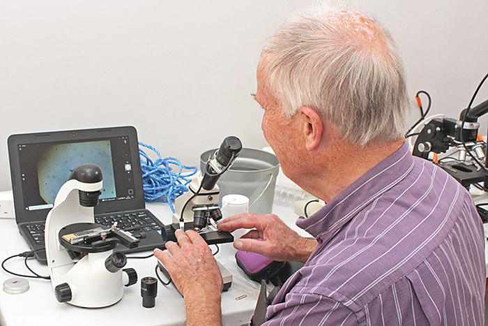
Mark Berry brought a simple monocular inverted microscope that is available branded as IQCrew or Amscope. He had attached a mechanical stage. He also brought a small monocular microscope with an eyepiece camera sending images via USB to his laptop computer.
 Mark Berry
New member Clare Blencowe and her husband Michael came for a chat and to see our equipment and specimens, and helped sample the millpond.
 Clare Blencowe, Robert Ratford and Stephen Durr
Stephen Durr brought his trinocular Leitz Dialux 20 EB with two nosepieces so that he could quickly switch between normal and phase contrast objectives. He used a Chinese inspection camera that records images on a card and displays them via HDMI on a monitor.
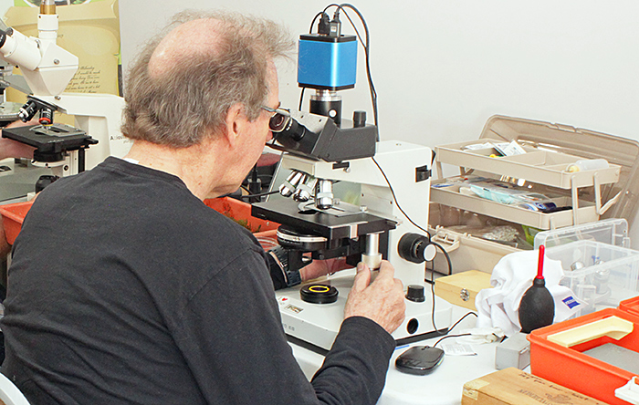 Stephen Durr
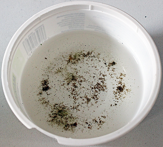
Stephen used a plankton net to collect a sample from the millpond
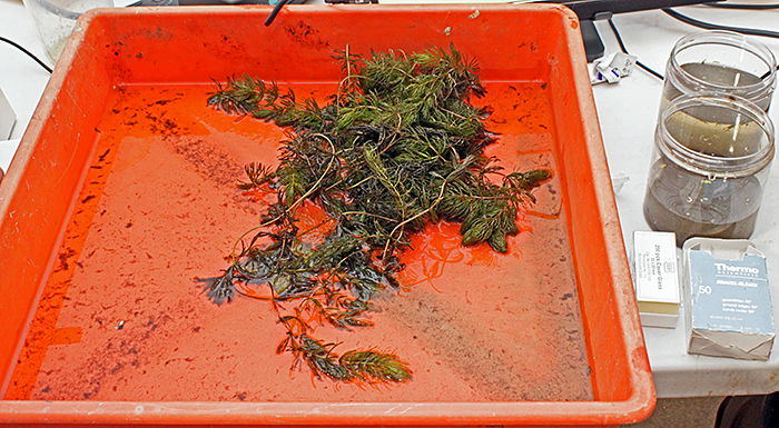 Stephen’s millpond sample
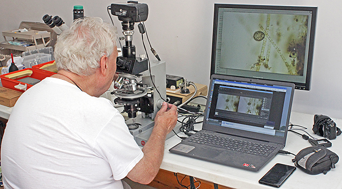
Neil Henry brought his monocular PZO compound microscope with 4×, 10×, 20× and 40× objectives and a YW5.0M eyepiece camera sending images to ToupView software on his laptop computer. He also brought a Veho Discovery USB digital microscope.
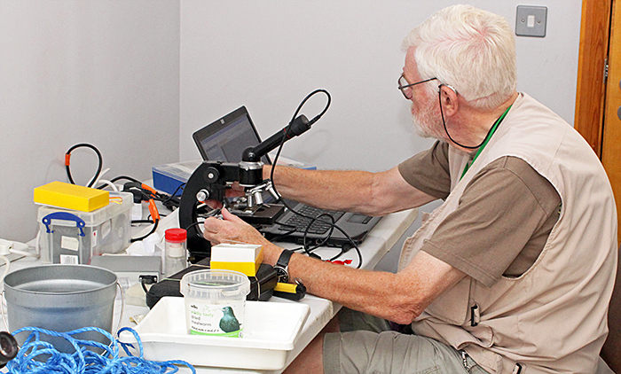 Neil Henry
Neil took a small bucket with a length of string tied to its handle to the millpond, and Clare Blencowe climbed down to the water’s edge to throw it in and pull it back.
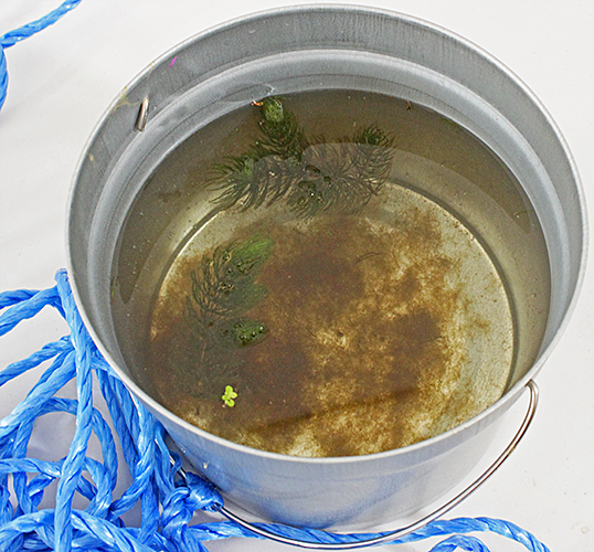 Neil and Clare’s millpond sample
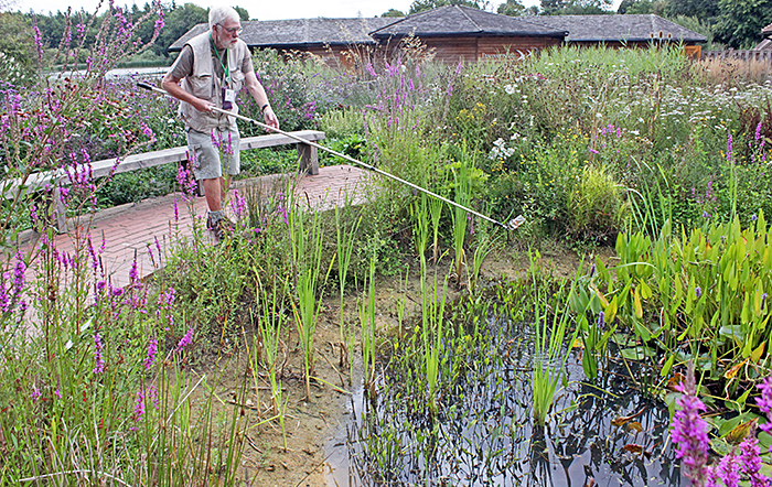 Neil Henry sampling the Shelley pond
Graham Matthews brought his familiar Leitz Dialux with 4×, 10×, 16×, 20× and 40× objectives, a circular stage and DIC. Lighting came via a dual fibre-optic gooseneck with both LED and electronic flash. He used his Canon EOS 500D digital SLR controlled by DSLR Remote Pro on his laptop computer. He used Helicon Focus to stack images for increased depth of field. He also brought a trinocular Wild M8 stereomicroscope with a transmitted-light stand.
 Graham Matthews
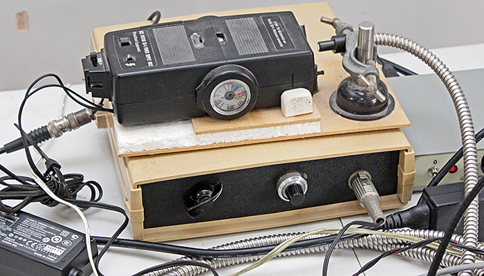 Graham’s modified electronic flash
Jacky McPherson didn’t want to carry a microscope on a long train journey, so she had arranged to borrow a grey trinocular Zeiss Standard microscope with a circular stage from Graham.
 Jacky McPherson
 Jacky McPherson sampling the reed bed
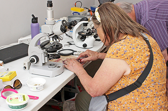
 Jacky’s reed bed sample
Jacky went for a walk to the walnut plantation, where she found a fruiting body of the saprotrophic sac fungus Daldinia concentrica on rotten wood. She brought it back and cut it open to reveal the concentric rings:
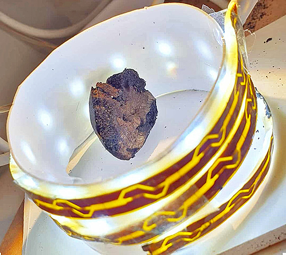 Daldinia concentrica (with Jacky’s LED strip lighting)
Robert Ratford brought a trinocular Zeiss Axiostar Plus with a set of Achrostigmat objectives from 5× to 100×. He used a smartphone held above an eyepieces to take photomicrographs. He also brought a grey Zeiss Jena binocular polarising microscope.
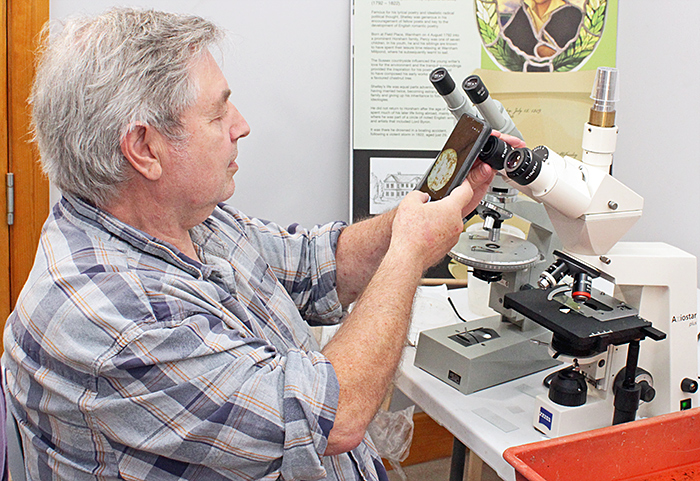 Robert Ratford
Robert also brought two small Elikliv digital microscopes that he used to show flowers as well specimens from the pond, including a mite and a budding green hydra.
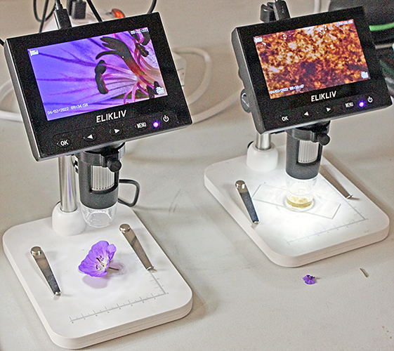 Digital microscopes
To hold a small sample of pond water, Robert used a holder made by cementing a short section of plastic tube to a slide:
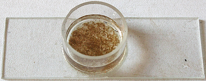 Specimen chamber
Photomicrographs
Here are some of the photomicrographs taken by Graham Matthews, Stephen Durr and Robert Ratford.
In the gallery below, click one of the small photos and you will see a larger photo with more information. On the large photo, click > or < at right and left to see the next and previous photos, or click ![]() at top right to start a slide show, or click
at top right to start a slide show, or click ![]() at top right to return to the gallery.
at top right to return to the gallery.
You can also use the right and left arrow keys on your keyboard to move between the large photos, and the Esc key to return to the gallery.
Gastrotrich and diatoms
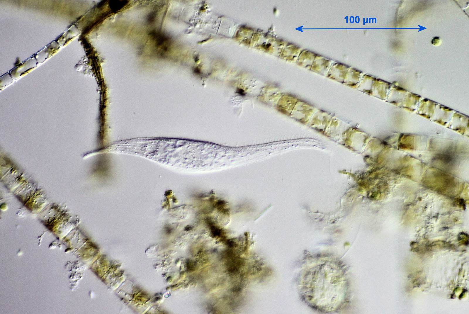
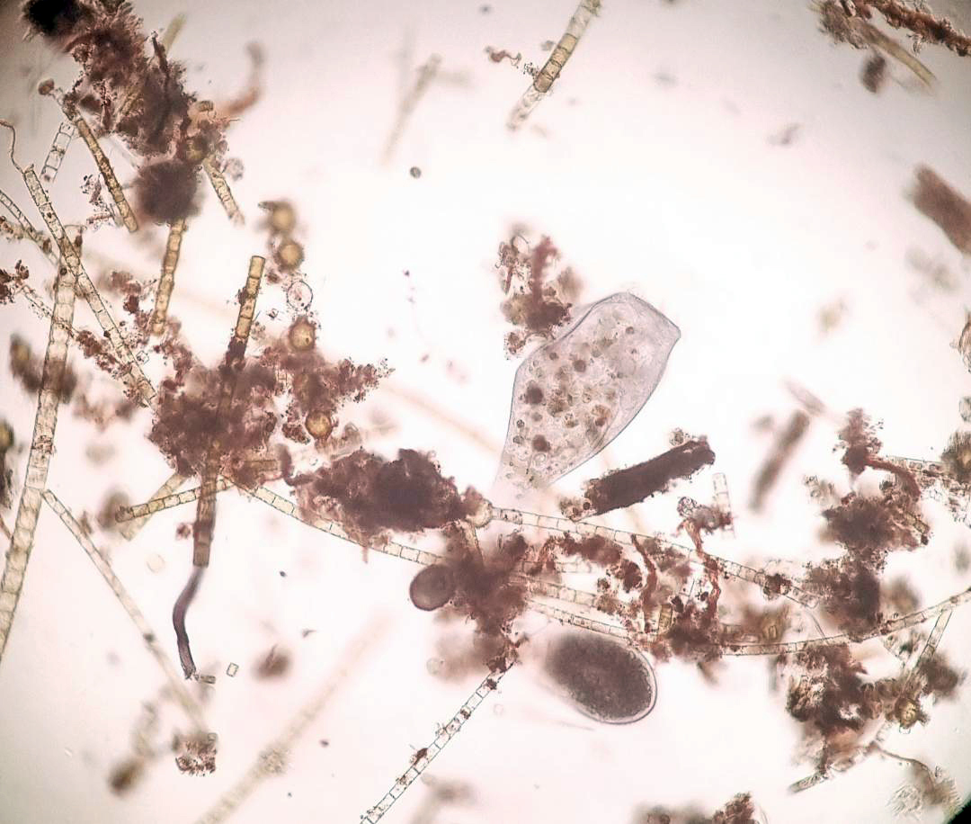
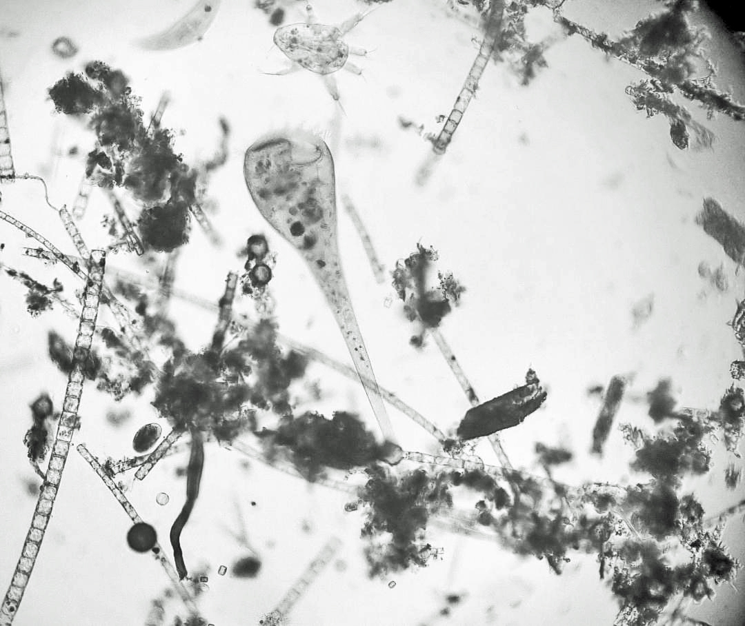
Protist: Litonotus sp.
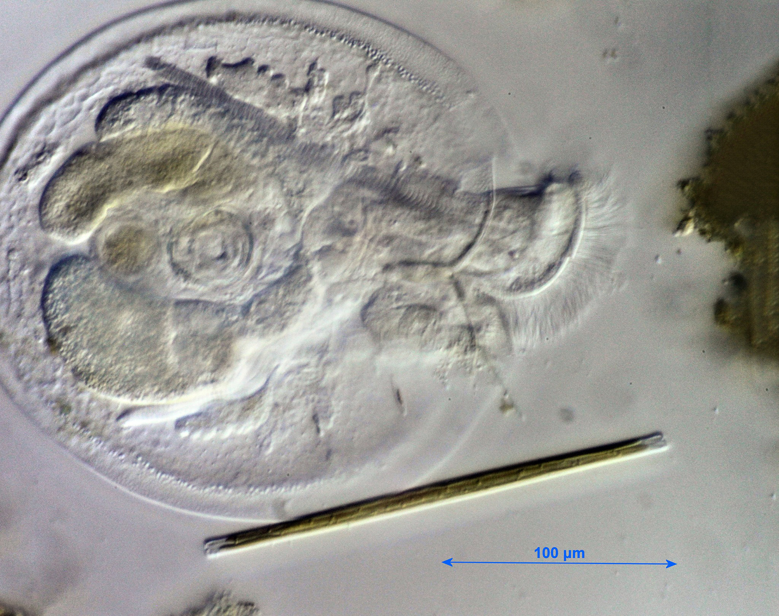
Rotifer: Testudinella patina

Rotifer: Testudinella patina
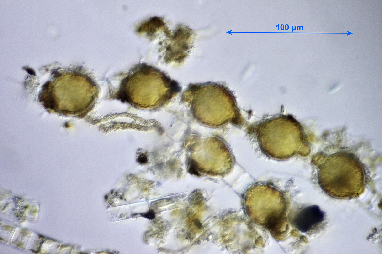
Algae (Chlorophyta): Oedogonium sp.
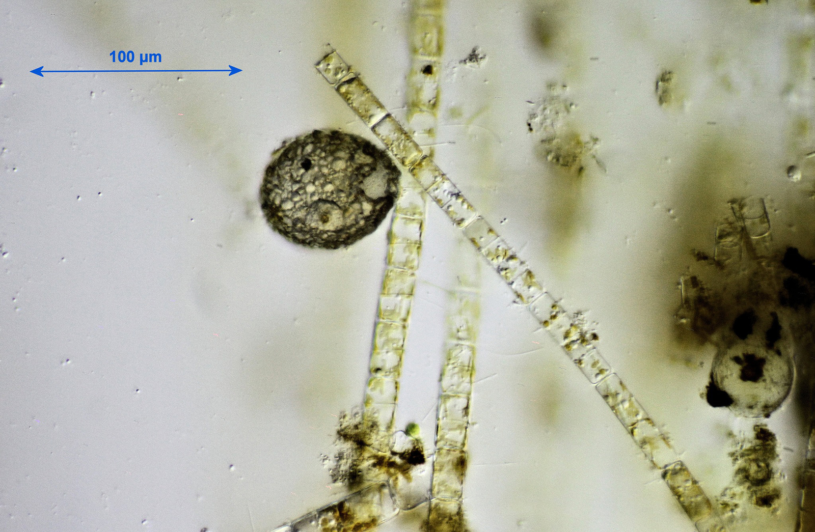
Amoeba test
Amoeba test
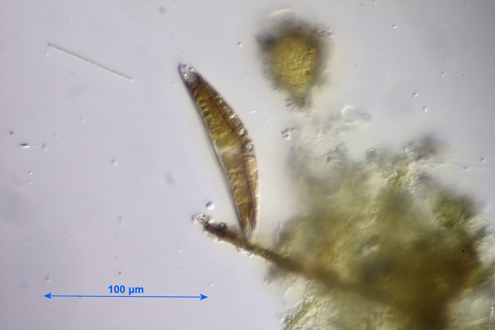
Diatom: Cymbella sp.
Diatom: Synedra sp. (?)
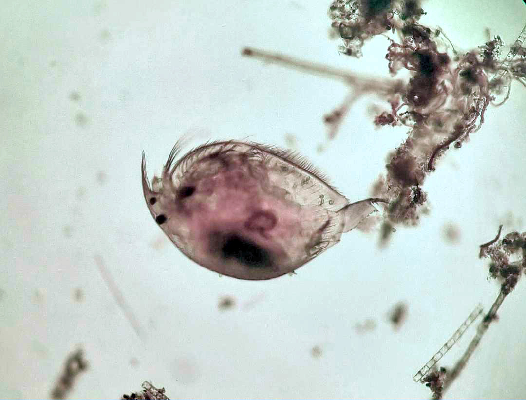
Scapholeberis mucronata
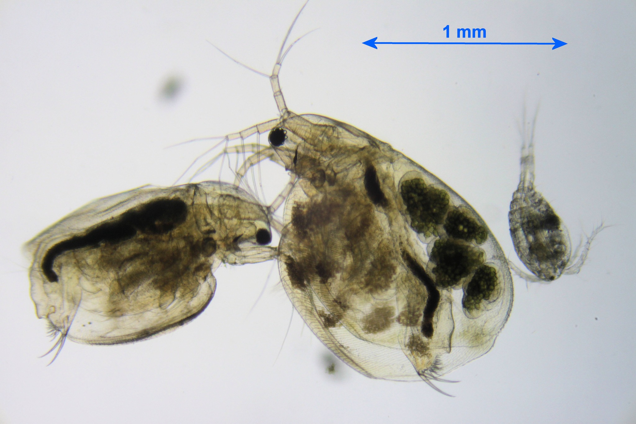
Simocephalus vetulus + cyclopean copepod
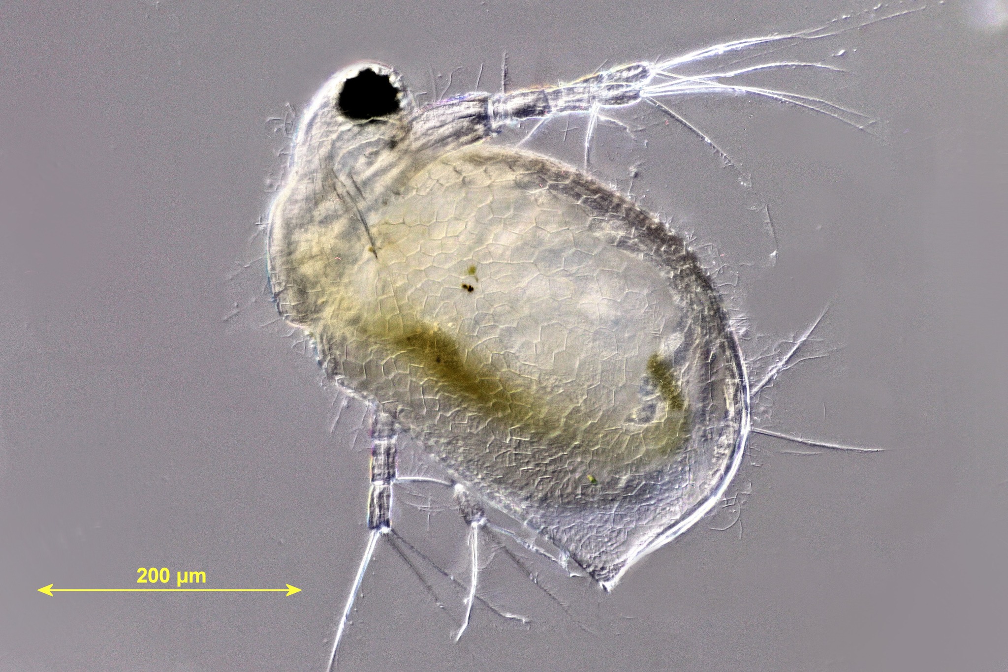
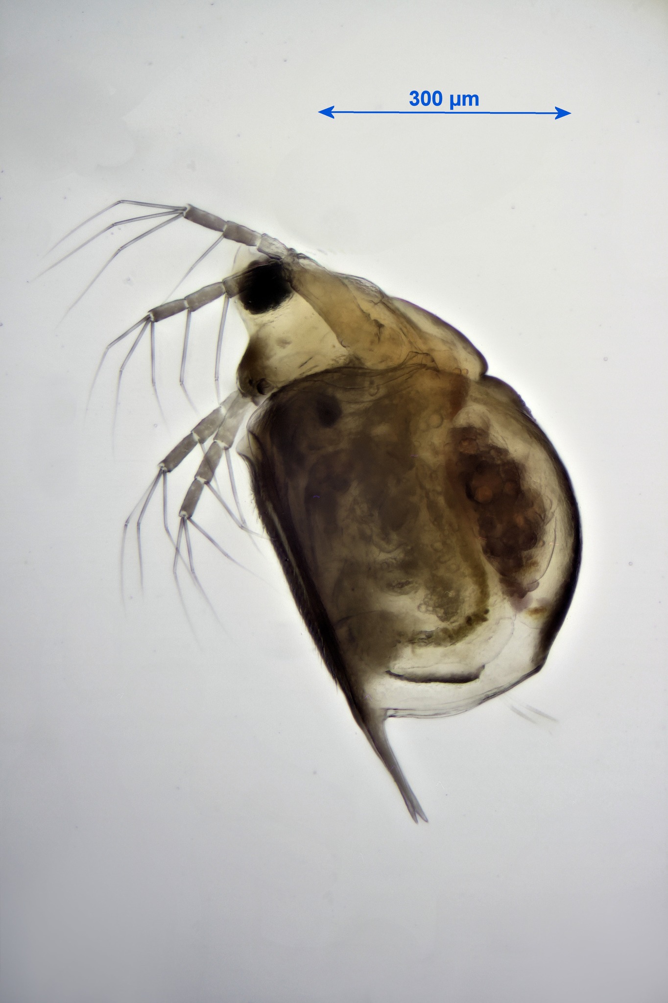
Ceriodaphnia reticulata
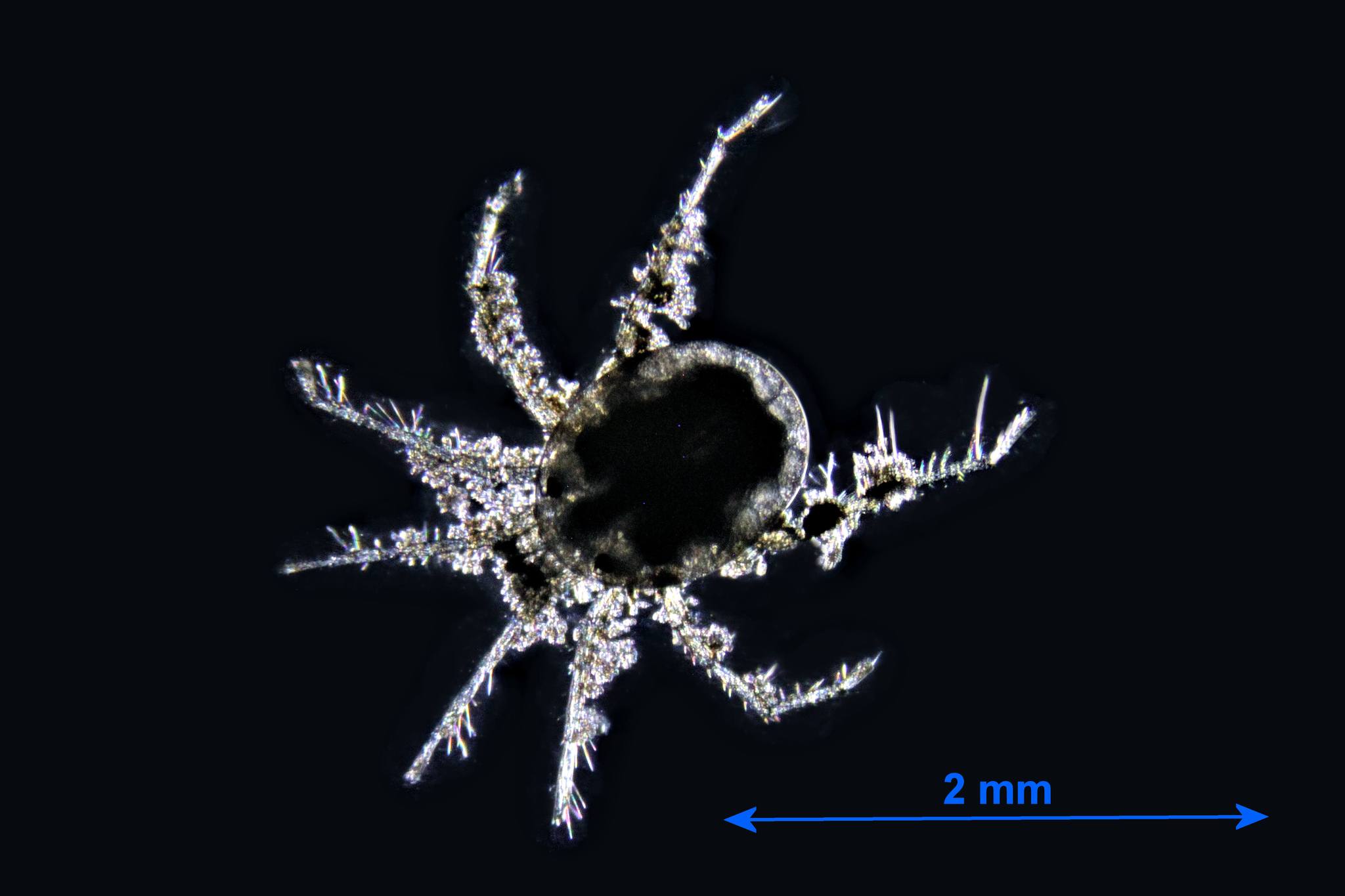
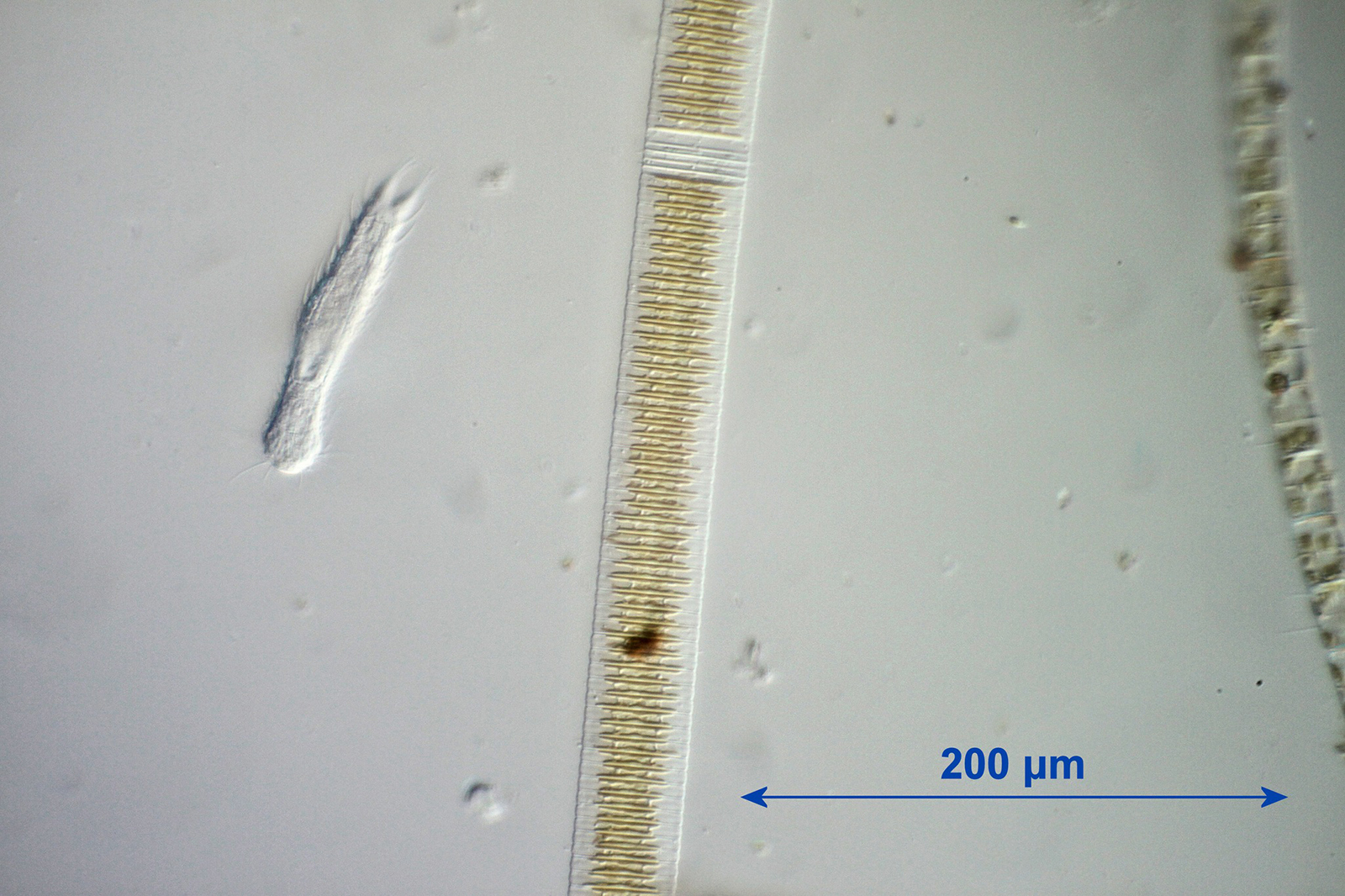
Hydracarina (freshwater mite)
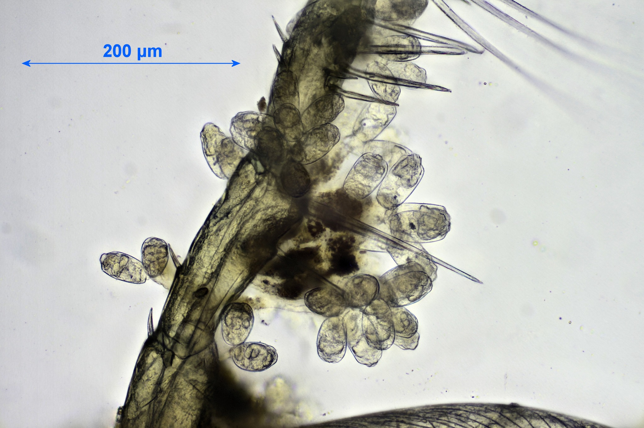
Epizootic peritrichs on Hydracarina
Nauplius larva
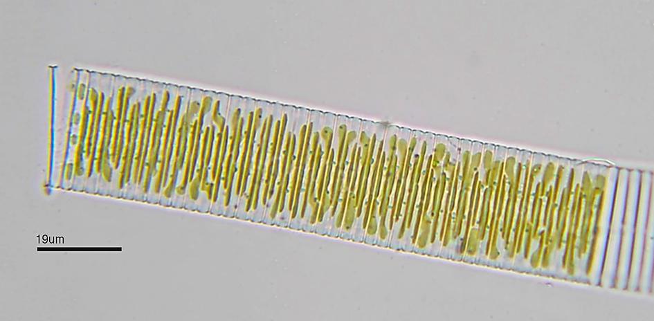
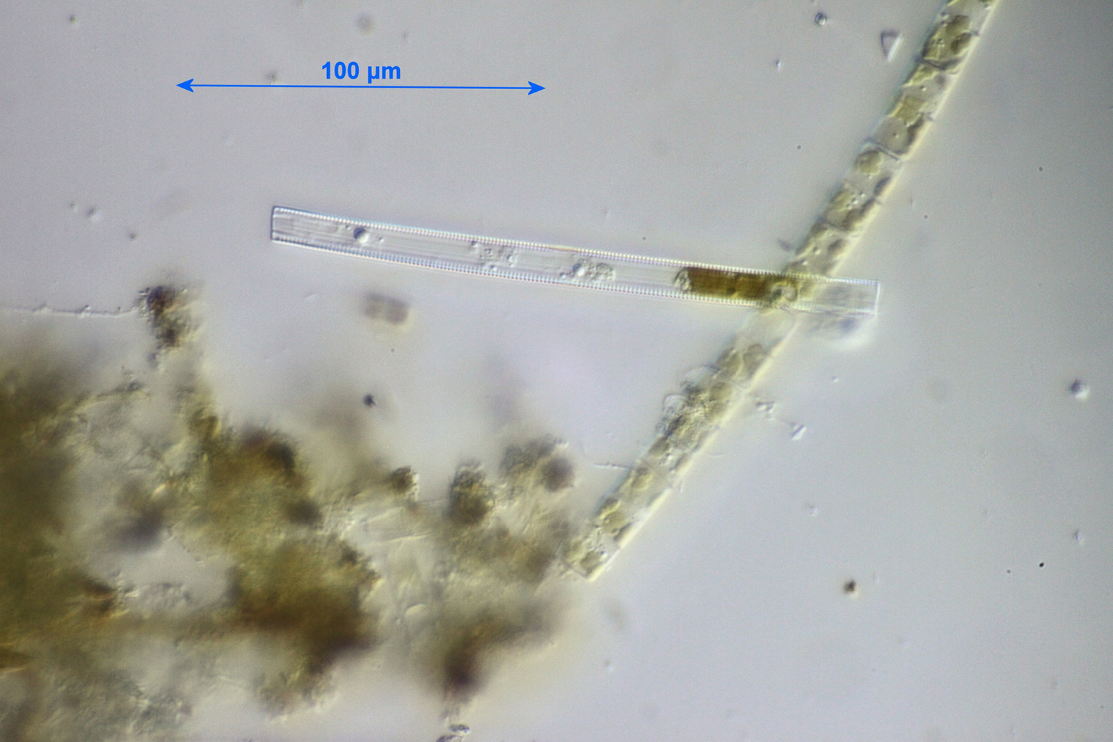
Diatom: Fragilaria sp.
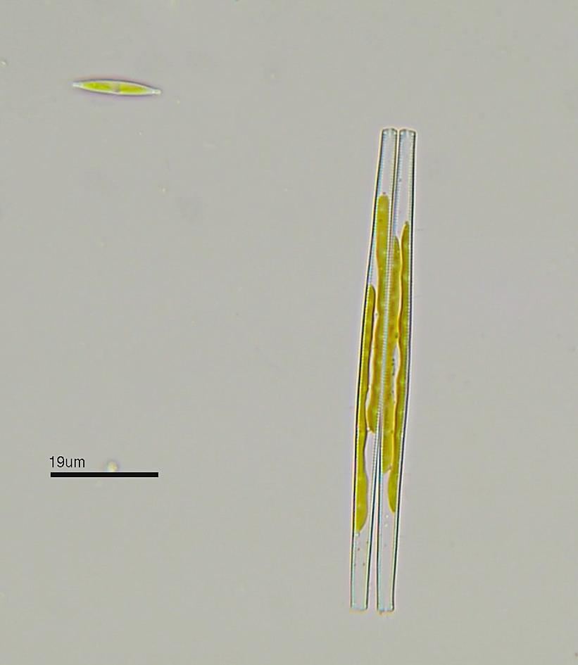
Diatom
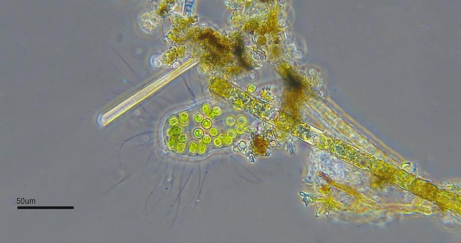
Algae: Apiocystis sp.
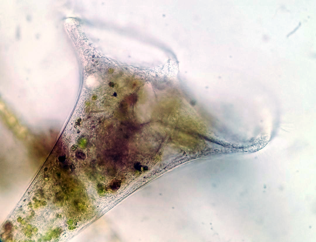
Ciliophora: Stentor sp.
Chydoridae
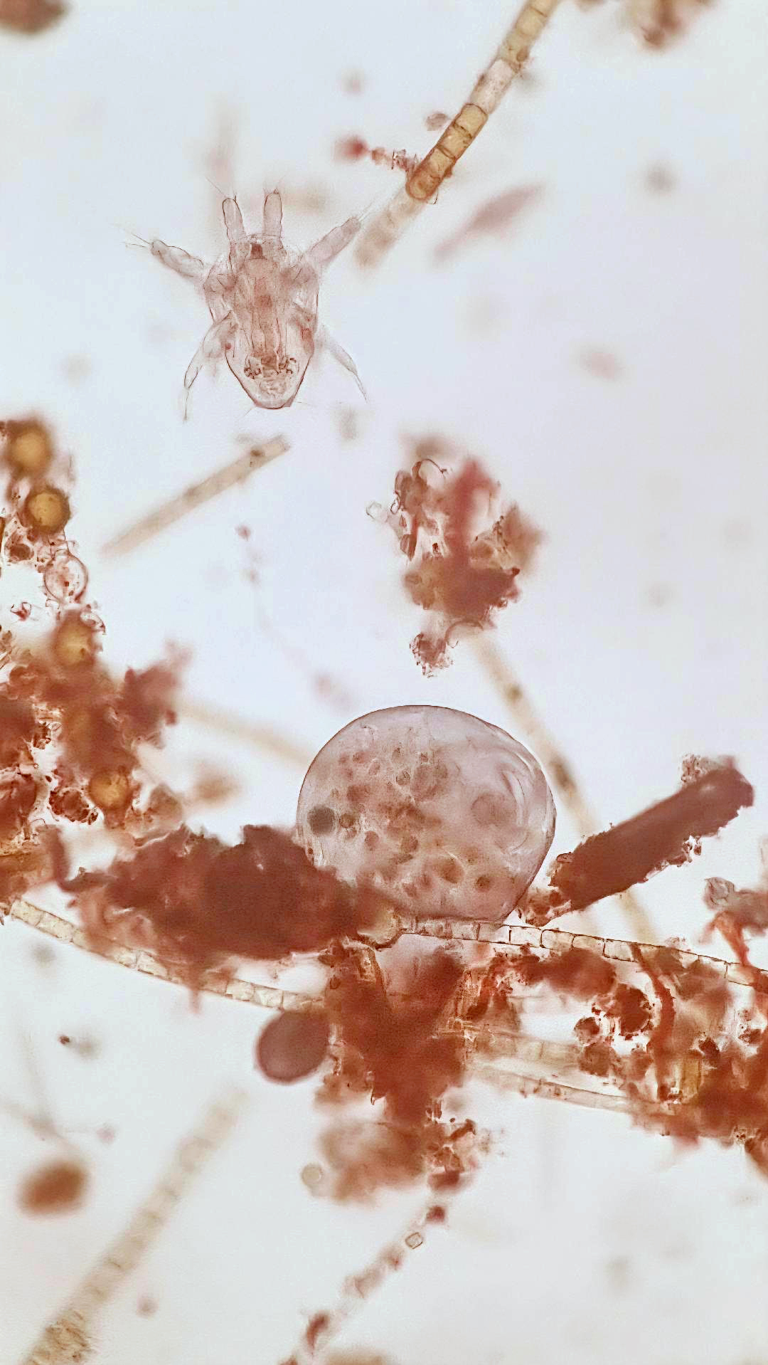
Nauplius larva
Rotifer
Rotifer
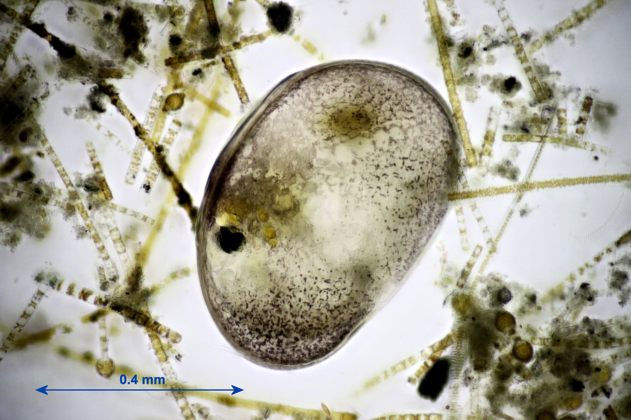
Ostracod
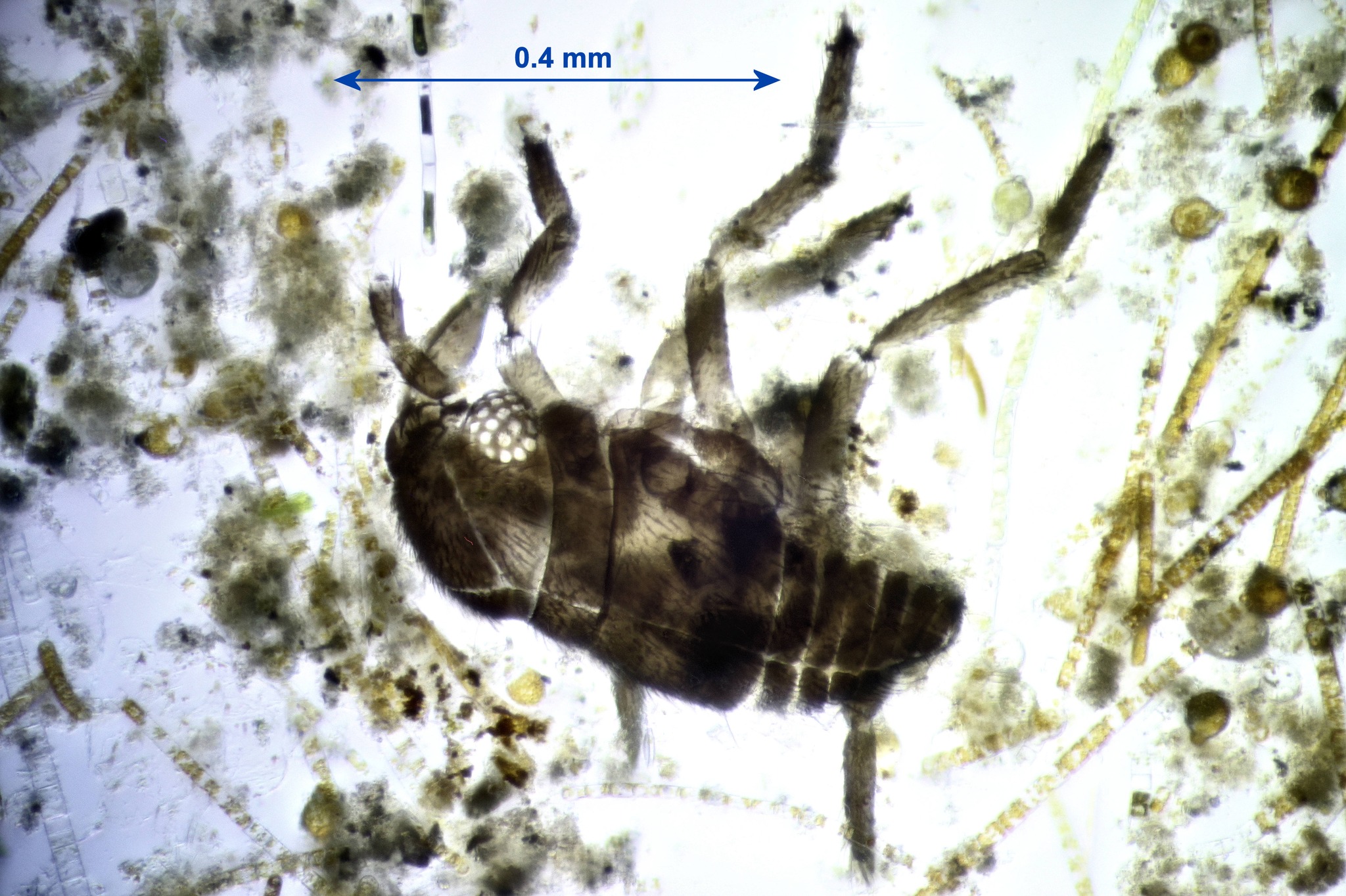
Insect
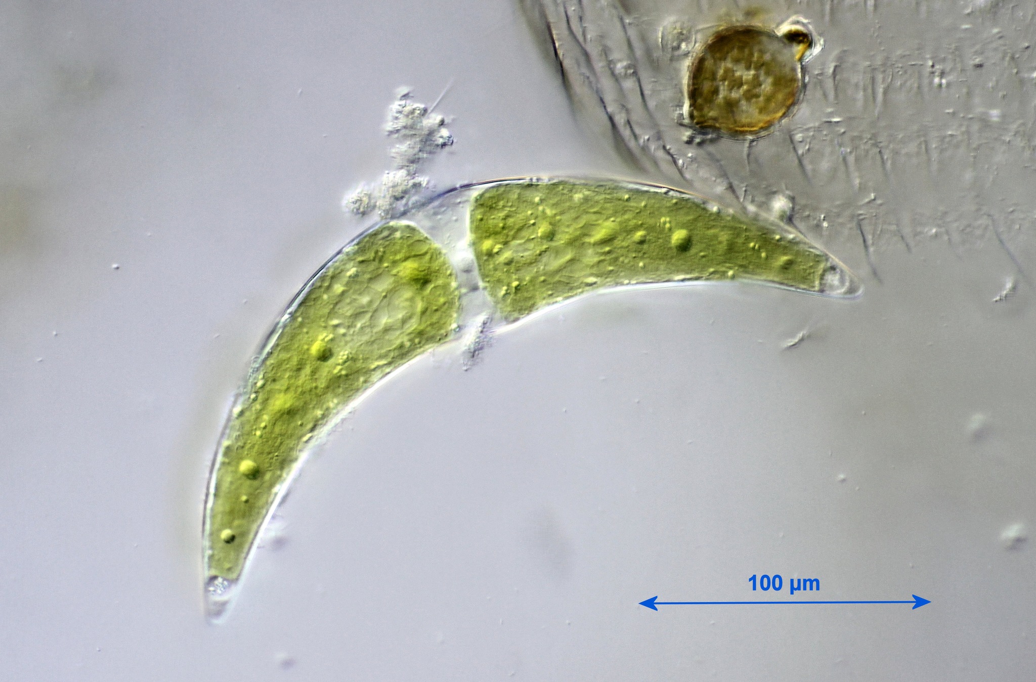
Desmid: Closterium sp.
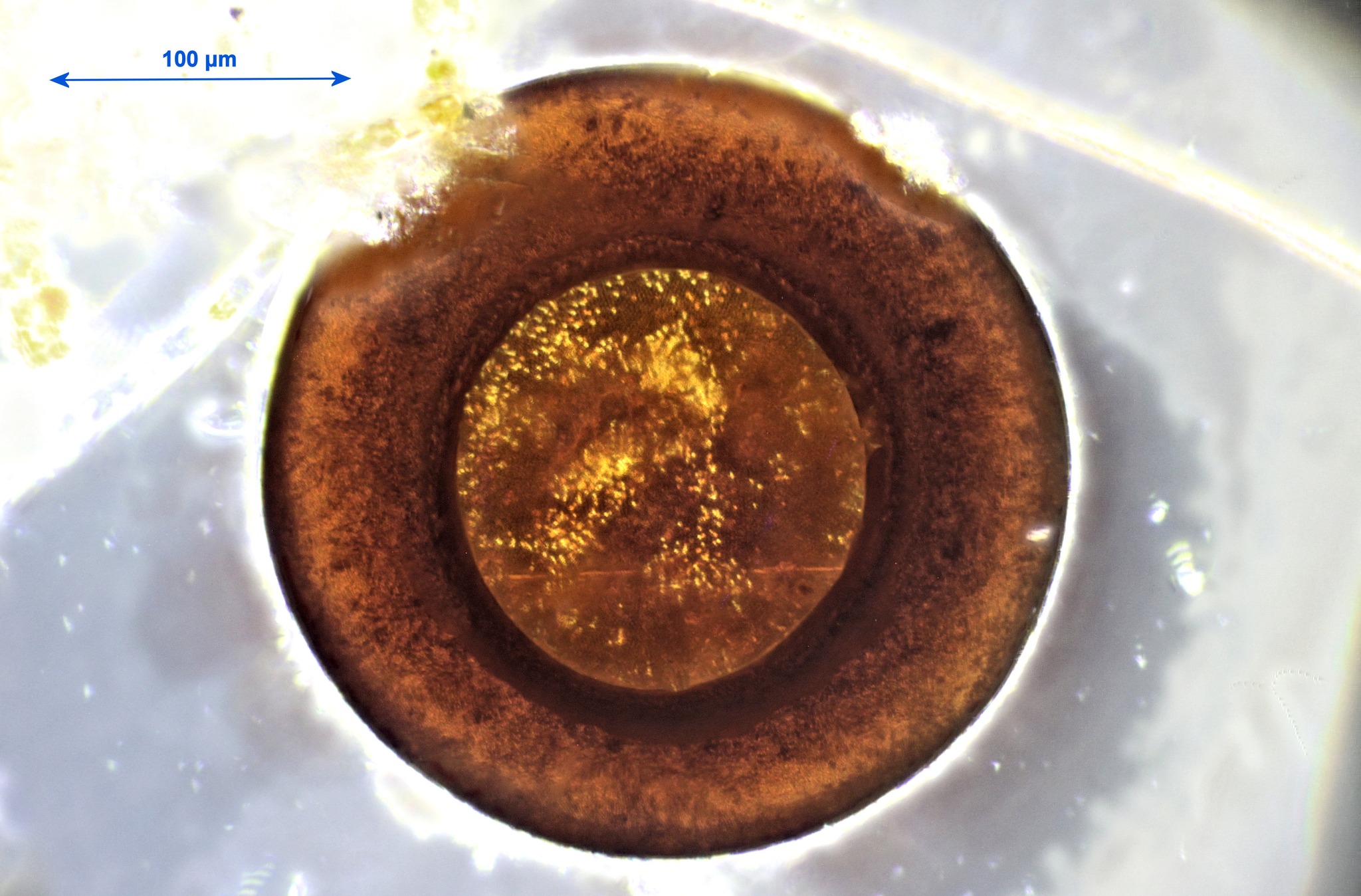
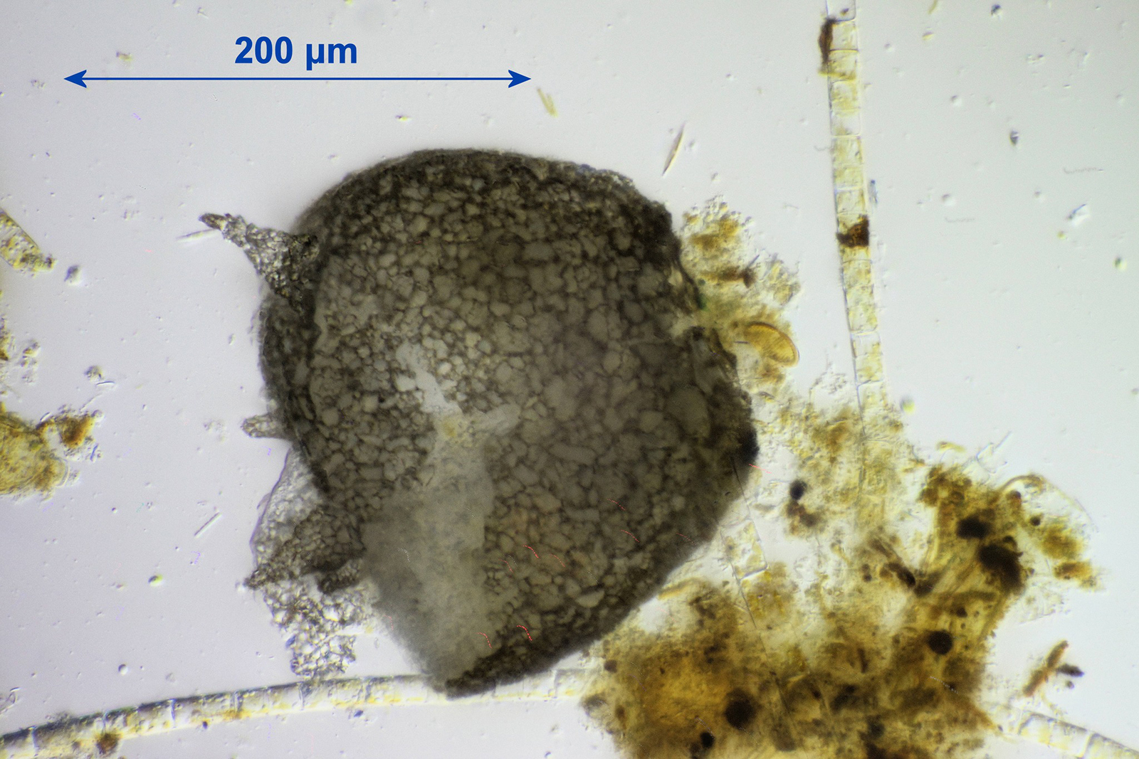
Amoeba test: Arcella sp .
Acknowledgements
Our thanks to the Friends of Warnham Local Nature Reserve for allowing us to collect specimens and to use the Discovery Hub, and to Graham Matthews for organising the excursion.
Report and most photographs by Alan Wood, photomicrographs by Stephen Durr, Graham Matthews and Robert Ratford.