

Down House Microscopy Weekend
Saturday 22nd and Sunday 23rd August 2026
This was our third visit to Down House in Kent, where Charles Darwin lived for 40 years. Quekett member Tim Fullick is one of the volunteers at Down House, and in the Discovery Suite he set up the highlight of the weekend, a display of brass microscopes similar to the ones that Darwin used.
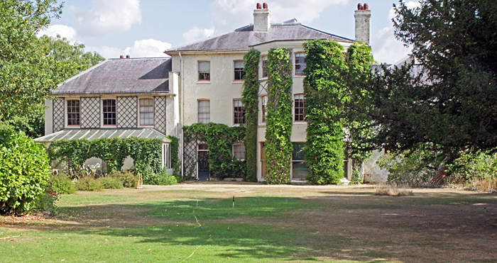 Down House
As in 2025, we had additional displays in the garden laboratory that is attached to the greenhouse and was built for Darwin. With more space (and with no fragile antiques), these extra displays were designed to be mostly hands-on for families.
Discovery Suite
Upstairs in Down House, we had displays of antique microscopes, carnivorous plants, rocks, wings and feathers.
Tim Fullick showed five antique brass microscopes, similar to the ones that Darwin used at various points in his career.
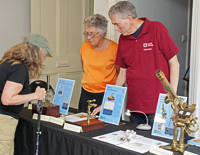 Tim Fullick
Tim had carefully set up the microscopes so that they were focused properly, and visitors were able to admire them and look through them, but were not allowed to touch them.
The Carl Zeiss Jena IVa was set up to view a slide of the body wall of an earthworm, showing circular and longitudinal muscle layers.
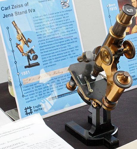 Carl Zeiss Jena IVa
The Smith & Beck Large Achromatic Compound Microscope was set up to view barnacle cirrus.
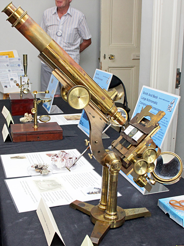 Smith & Beck Large Achromatic Compound
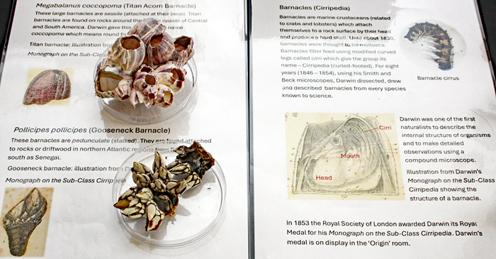 Barnacles
The Hartnack Nouveau Petit Modèle was set up to view the head of a Drosera (sundew) tentacle.
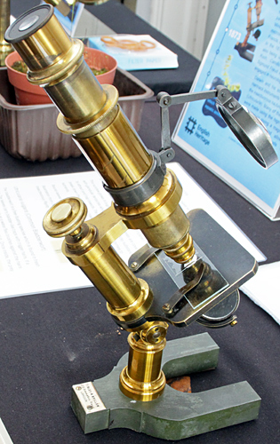 Hartnack Nouveau Petit Modèle
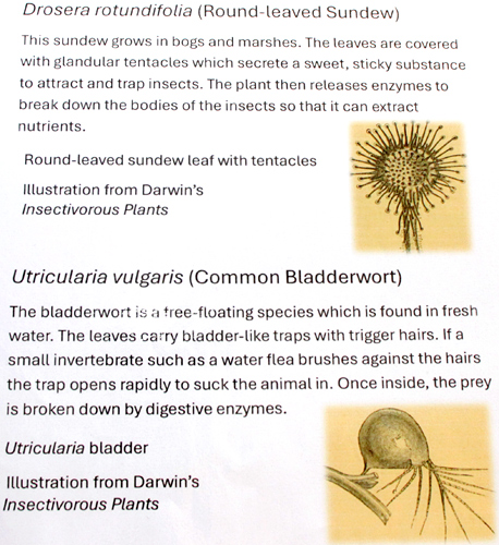 Insectivorous plants
The Cary-Gould Box-Mounted Microscope was set up to view zooids of Flustra (a bryozoan).
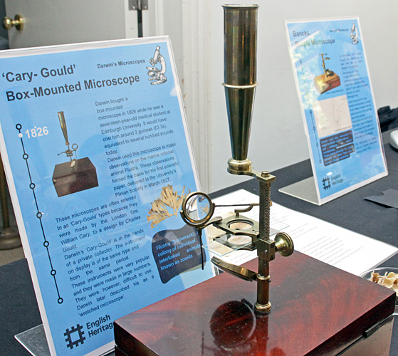 Cary-Gould Box-Mounted Microscope
The Bancks Simple Microscope was set up view waterfleas (Daphnia).
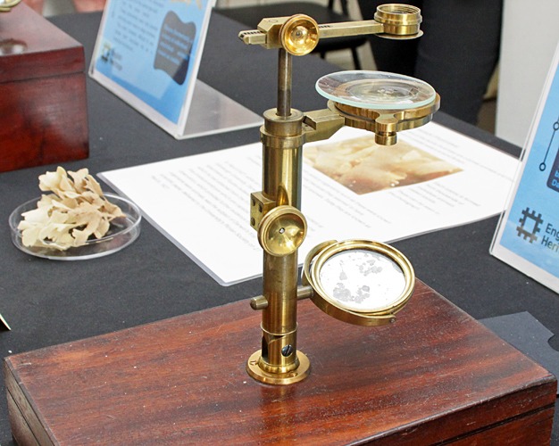 Bancks Simple Microscope
Lisa Ashby’s display this year was about wings, with examples and illustrations of wings of bats, birds, beetles, butterflies and moths. Lisa provided a small stereo microscope and two digital microscopes with small built-in screens so that we could examine bird feathers and parts of butterfly wings mounted in holders for 35mm slides.
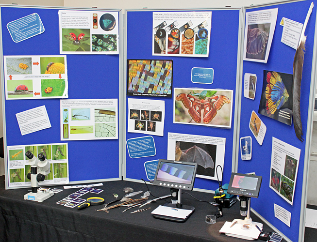 Lisa Ashby’s display
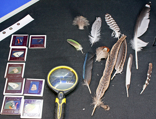 Butterfly wings and feathers
Joan Bingley brought some carnivorous plants (bladderworts, pitcher plants and sundews) as well as prepared slides. She provided a digital microscope with a small built-in screen, a binocular compound microscope and two stereo microscopes so that we could view the slides and various aspects of the plants.
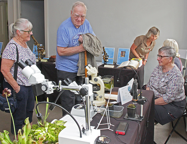 Joan Bingley (right)
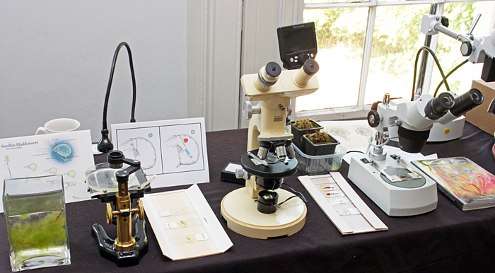 Joan Bingley’s display
Pam Hamer’s display was designed to inform us about Darwin’s interest in geology, including the different origins and compositions of rocks, and the presence of fossils and microfossils.
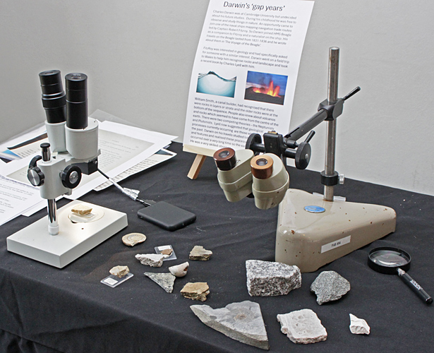 Pam Hamer’s display
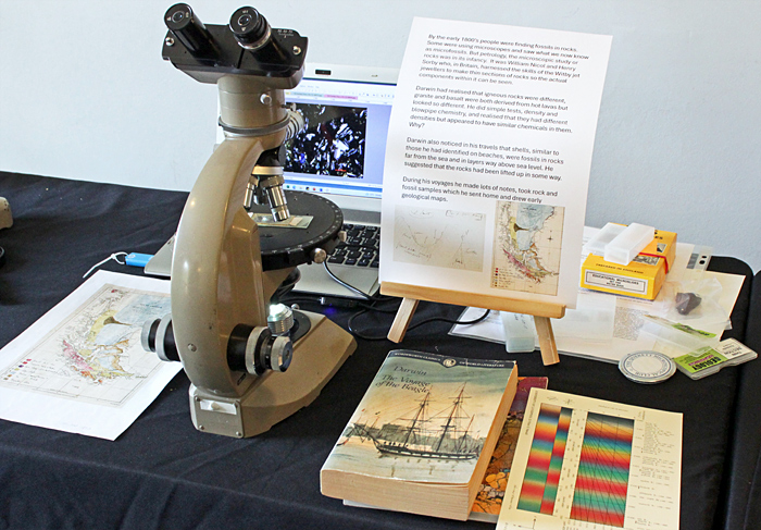 Pam Hamer’s display
Martin Parnham brought a small digital microscope with a built-in screen, and a modern trinocular compound microscope with a camera linked to a monitor. His specimens included eggs of brine shrimps that were hatching, and barnacles.
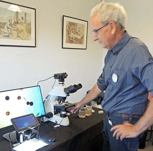 Martin Parnham [by Jacky McPherson]
Nigel Williams brought some slides made by Klaus Kemp, including one with several species of diatoms and two made by arranging scales from butterfly wings into a peacock and a vase of flowers.
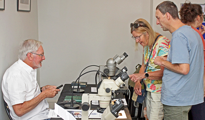 Nigel Williams
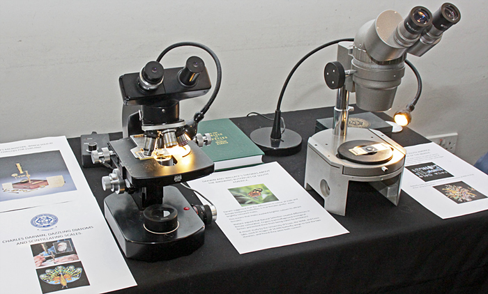 Nigel Williams’ display
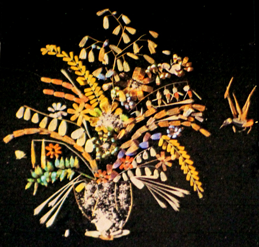 Klaus Kemp arrangement of butterfly scales
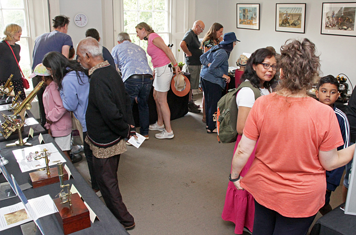 Visitors
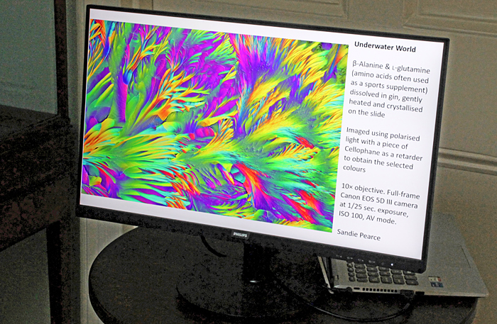
At times, visitors were asked to queue outside the room to see our exhibits. To keep them entertained, we used a slide show of photomicrographs taken by Quekett members:
 Slide show
Garden laboratory
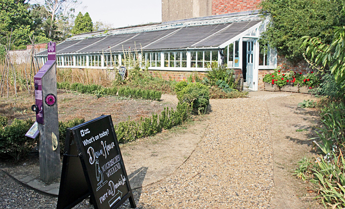
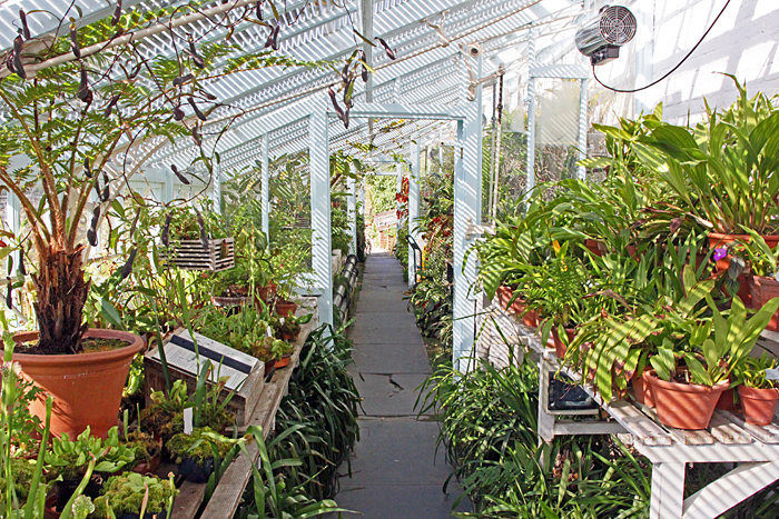
The garden laboratory is down a few steps inside the greenhouse, and there is also step-free access via the garden. The permanent attraction in the laboratory is usually an observation beehive, but this year the bees deserted it. Quekett members set up their microscopes so that visitors could view specimens from the garden and elsewhere
 Greenhouse
 Inside the greenhouse
 Pitcher plants
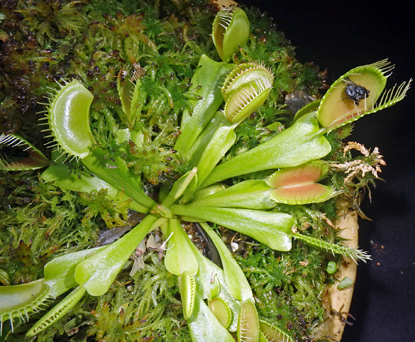 Venus flytrap [by Pam Hamer]
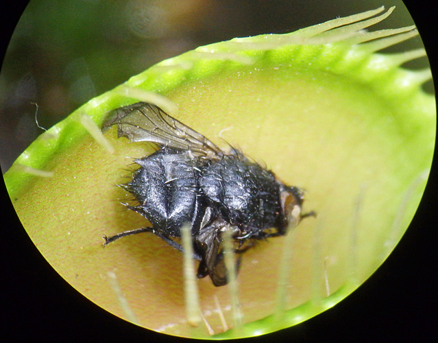 Fly in Venus flytrap [by Pam Hamer]
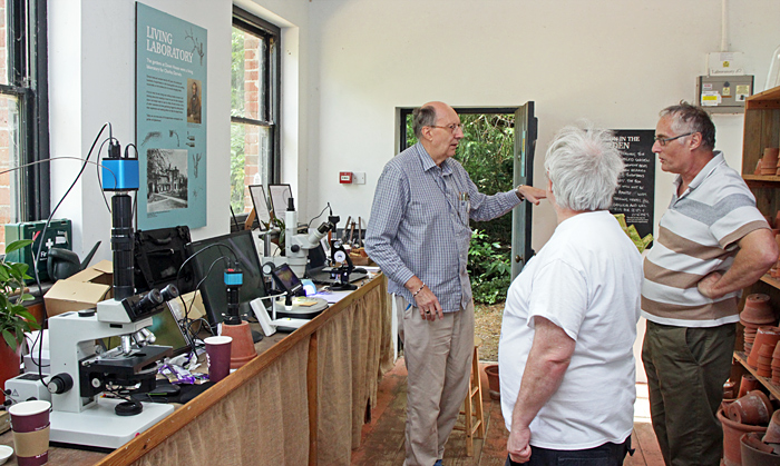 Garden laboratory (Chris Thomas, Robert Ratford and Laurence Peacock)
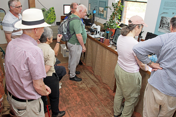 Visitors in the garden laboratory
Stephen Durr brought his trinocular Leitz Dialux 20 EB compound microscope (equipped for bright field and phase contrast) with a camera sending images to a monitor. He used it to show images of pond life, including the colonial green alga Pediastrum.
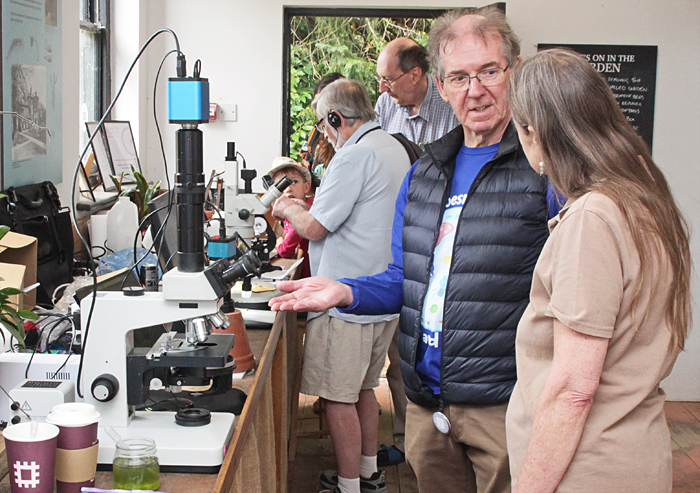 Stephen Durr
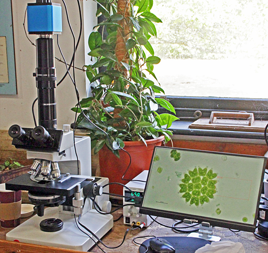 Stephen Durr’s display
Pam Hamer used a simple Lomo compound microscope to show slides of pollen and bee parts.
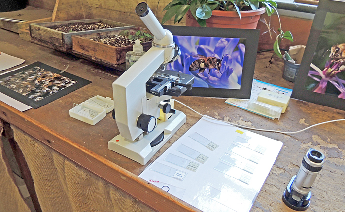 Photographs and slides of bees and pollen [by Jacky McPherson]
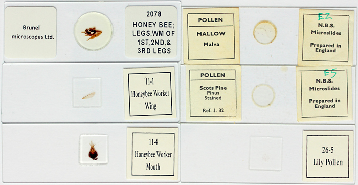 Commercial slides of bee parts and pollen
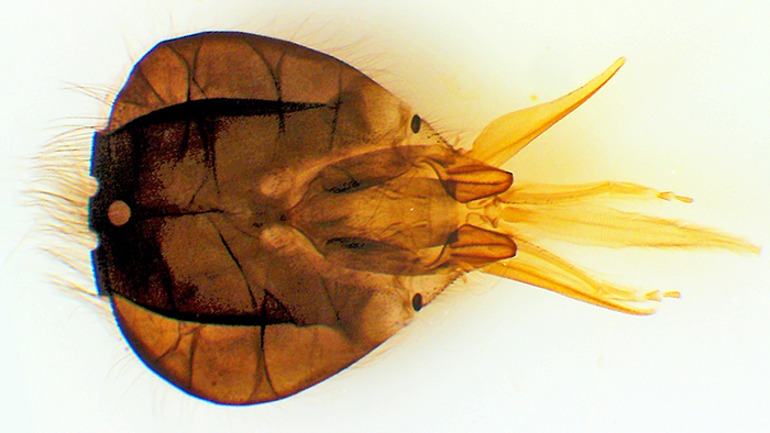 Head of honeybee [by Pam Hamer]
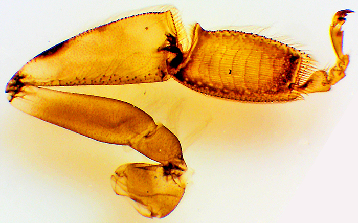 Hind leg of honeybee [by Pam Hamer]
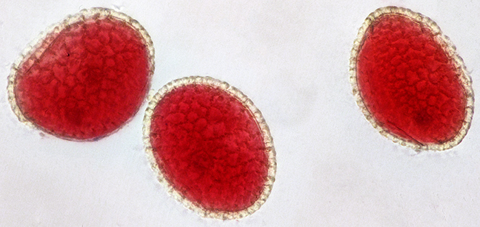 Stained grains of lily pollen
Jonathan Palmer showed some mounted photographs of bees.
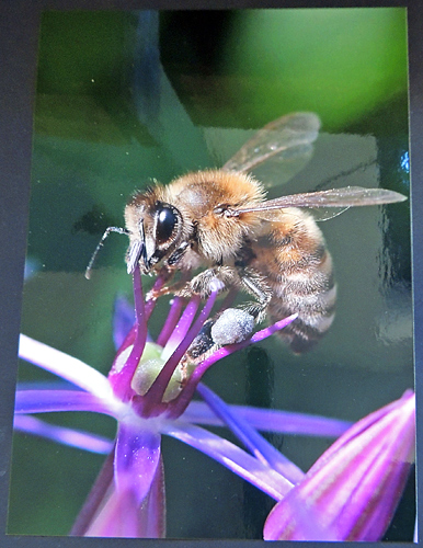 Photograph of bee
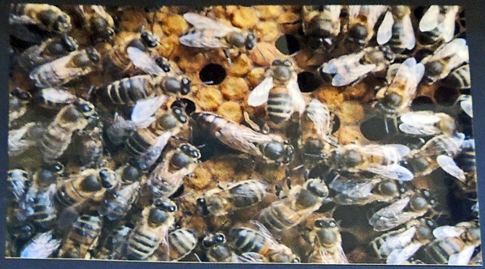 Photograph of bees
Laurence Peacock used a digital microscope to show the scales and other details of butterflies. He also used a compound microscope linked to a laptop to show live freshwater specimens in a Petri dish, and slides including bee parts.
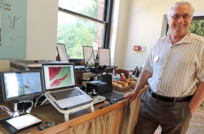 Laurence Peacock [by Jacky McPherson]
Robert Ratford used an inspection camera linked to a monitor, a digital microscope with a small built-in screen and a stereo microscope to show specimens from the greenhouse and the gardens. The specimens included a small yellow fungus known as the “flowerpot parasite” Leucocoprinus birnbaumii, and mosquito larvae.
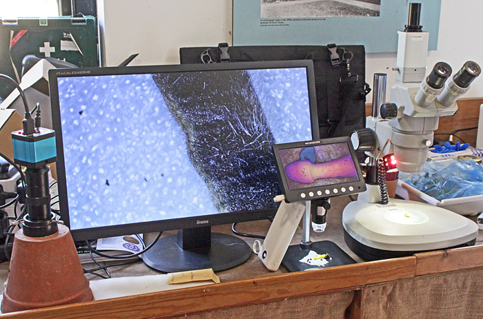 Robert Ratford’s display
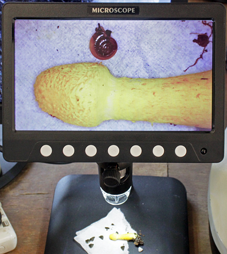 Flowerpot parasite (and a tiny snail) from the greenhouse
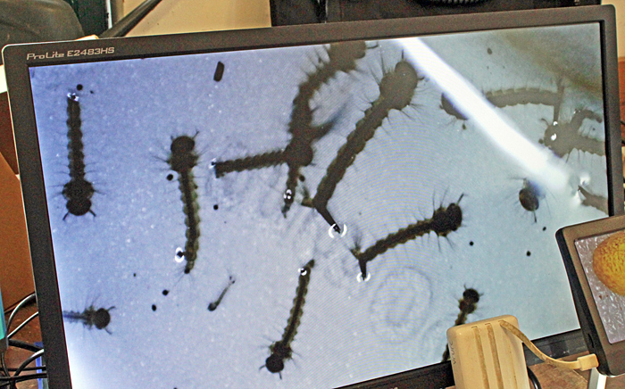 Mosquito larvae from a water butt
Chris Thomas brought a monocular microscopes from the 1960s, brought up-to-date with LED illumination and fitted with a digital C-mount camera instead of an eyepiece, so that images could be seen and captured on a laptop computer.
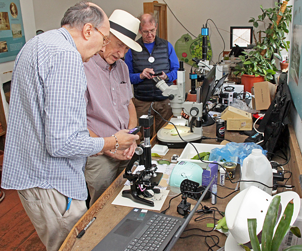 Chris Thomas
Chris encouraged visitors to cut a square (15–20 mm) from a fresh leaf and press it onto a drop of Vida Rosa UV resin on a slide. The leaf is then covered with non-stick baking paper, another slide is placed on top, and the slides are clamped together with two small metal clips. Then the assembly is put into a UV lamp that is designed for setting nail polish, and exposed for 60 seconds. This hardens and dries the resin. After separating the parts and peeling off the leaf, the slide is ready to be labelled and examined under a microscope so that the stomata and other features can be seen.
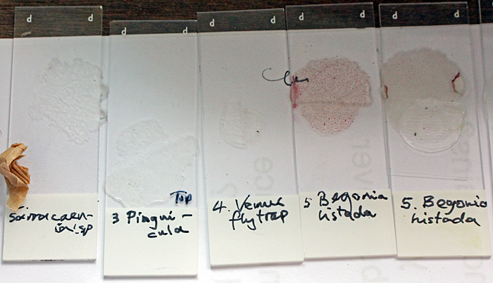 Leaf prints
Acknowledgements
Massive thanks to the English Heritage volunteers who marshalled the visitors so that we were not swamped, while encouraging them to see and interact with our displays in the Discovery Suite and the garden laboratory. English Heritage recorded over 400 visitors to Down House over the weekend.
Report and most photographs by Alan Wood