

It’s a small world after all
Wednesday 23rd October 2019
These two family-friendly microscopy workshop sessions aimed at 11-14 year olds (with their parents) were organised by the Museum of Life Sciences of King’s College London and the Hunterian Museum of the Royal College of Surgeons and held in the Museum of Life Sciences in the Hodgkin Building on Guy’s Campus in London. The workshop was led by Dennis Fullwood, assisted by Quekett members Danny Ferri, Jacky McPherson, Paul Smith and Alan Wood, and Jill Sales (Museum of Life Sciences) and Hayley Kruger (Royal College of Surgeons), as part of the Club’s outreach programme.
Dennis Fullwood introduced the two sessions with a short talk covering the early history of microscopes and a comparison of compound microscopes and stereomicroscopes. You can find out more about the relative advantages of stereo and compound microscopes in the Starting with microscopes section of the Quekett website.
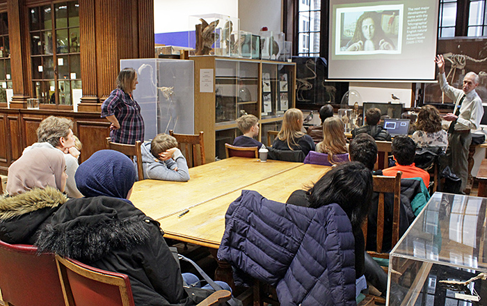 Dennis Fullwood’s introduction (first session)
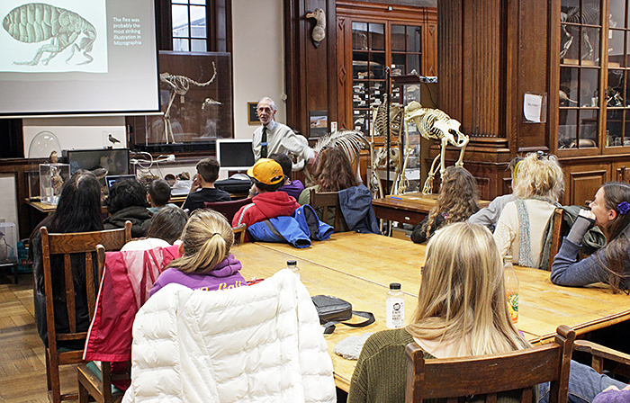 Dennis Fullwood’s introduction (second session)
After the introduction, the participants were split into 2 groups to take turns at making a slide of the jumping leg of a locust and examining a range of slides and specimens using compound microscopes, stereomicroscopes and a digital microscope.
The microscopes were 2 brass Watson compound microscopes made around 1900, a Nikon Labophot compound microscope from the 1970s, an Olympus SZ4045 stereomicroscope from the 1990s, 2 simple modern stereomicroscopes, a digital microscope with a small built-in screen (KKmoon Digital USB Microscope), an inspection camera sending an image to a monitor, and a replica Leeuwenhoek microscope.
Some of the microscopes had built-in illumination, and for the others we used JANSJÖ lamps from IKEA. The mains-powered JANSJÖ lamps seem to have been replaced by the less flexible NÄVLINGE lamps.
Specimens included a large number of microscope slides for viewing with the compound microscopes, and insects in amber, star sand, a feather, a postage stamp and a printed circuit board for viewing with the stereomicroscopes. One grain of star sand was glued to the specimen holder of the replica Leeuwenhoek microscope.
The Quekett members explained how to focus the compound microscopes without breaking the slides, by starting with the objective close to the slide and then using the coarse focus to raise it until the specimen comes into focus. We also showed them how to put slides into the mechanical stage so that they can easily be moved left and right and backwards and forwards. For the stereomicroscopes, we showed the participants how to adjust the distance between the eyepieces so that they could see a 3-dimensional image.
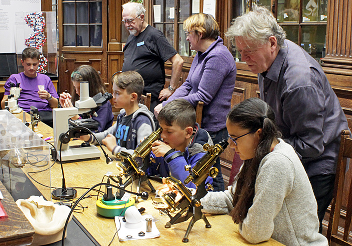
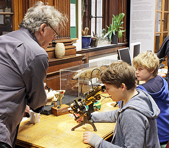 Danny Ferri and 2 young participants with Watson brass microscopes
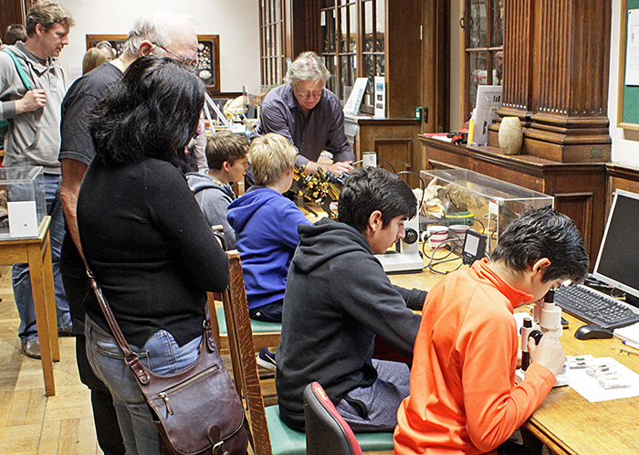
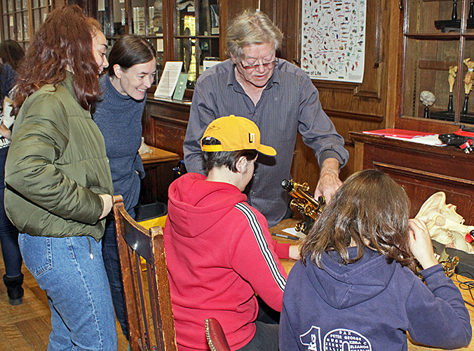 Danny Ferri with parents and children
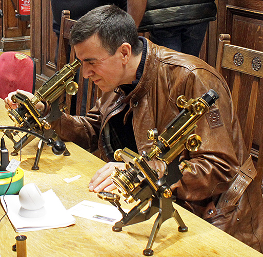 Parent with Watson brass microscopes
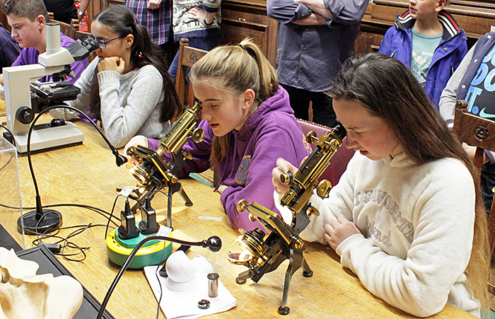
 Young participants with microscopes
 Paul Smith, Danny Ferri and participants
 Young participants with microscopes
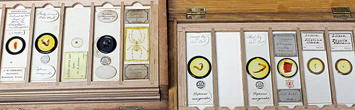 Microscope slides
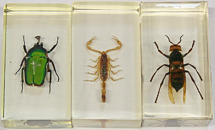 Insects and scorpion in resin
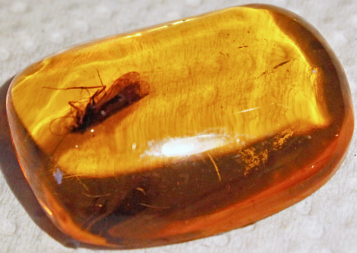 Insect in amber
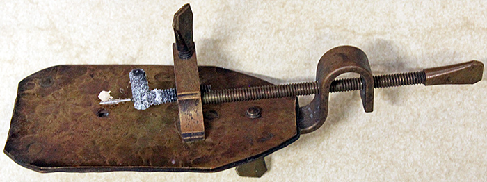 Replica Leeuwenhoek microscope, with a grain of star sand
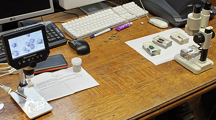 Digital microscope and small stereomicroscope
We were not able to use glass microscope slides for the participants to take away, so Dennis provided 3″×1″ card slides to which he had glued the bottom part of coin capsules. The glue was clearly visible through the clear base of the capsules, so Dennis provided circles of white paper that the participants could glue onto the base of the capsules. Dennis provided a jar of young locusts and lots of tweezers and scissors so that the participants could remove a jumping leg and glue it to the white paper. They were able to use the inspection microscope or a stereomicroscope to help them remove a leg. The slides were then set aside to dry before fitting the top of the coin capsules.
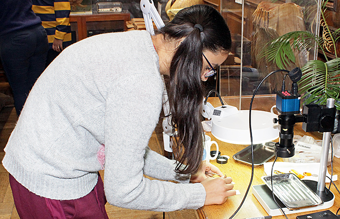 Making a slide
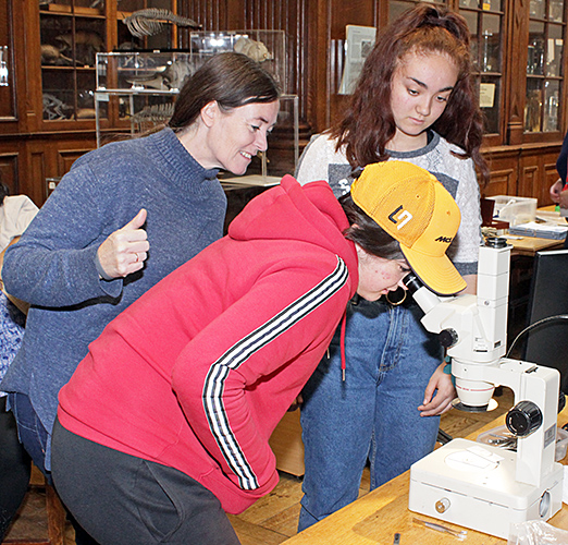 Checking a slide under a stereomicroscope
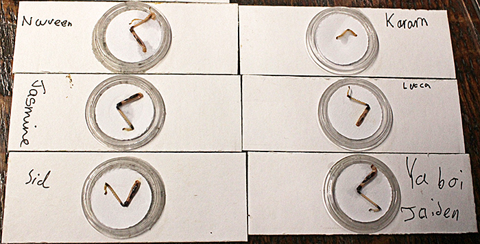 Slides of locust legs
For anyone wanting to make their own slides, Brunel Microscopes and Magnacol sell slides, coverslips, mountants, stains, ringing cements and several chemicals.
Coin capsules are easy to find on Amazon and eBay, and the idea of using them for dry mounts came from another Quekett member, Colin Lamb. The sizes quoted are the internal diameter, so 21 mm is the largest that will fit on a standard slide.
Before and after the workshop sessions, the participants were able to examine the wide range of exhibits in the Museum of Life Sciences.
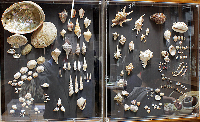 Display of shells
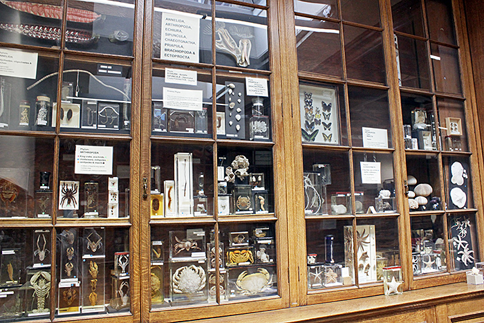 Display cabinets
The team members were kept very busy fielding questions and enquiries from participants and their parents. Popular subjects included slide making techniques, acquisition of microscopes and equipment, and attendance at other Quekett events.
Hayley has analysed the evaluation forms and they were most encouraging, with 70% saying the event was excellent. The most frequent answers to “What did you most like about the event?” were the chance to examine interesting slides with both Victorian and modern microscopes, and the slide making. There were also many favourable comments about the enthusiasm and expertise of our team.
Report and photographs by Alan Wood